
Hello again. In the previous lesson, you learned that proteins are polymers made from amino acids joined by peptide bonds. This lesson takes the next essential step: a protein is not useful merely because it is a long amino-acid chain. Its amino-acid sequence causes it to fold into a particular three-dimensional shape, and that shape allows it to carry out a particular biological function.
By the end, you should be able to explain the full chain of cause and effect:
From a polypeptide chain to a working protein
A polypeptide is a chain of amino acids joined by peptide bonds. Amino acids all have a similar basic structure, but each type has a different R group (also called a side chain). It is these R groups that make one amino acid different from another.
For example, an R group may be:
- hydrophobic: repelled by water;
- hydrophilic: attracted to water;
- positively or negatively charged;
- able to form particular bonds with other R groups.
The primary structure of a protein is the exact order of amino acids in its polypeptide chain. Changing the order changes which R groups occur at each point along the chain.
This matters because the R groups interact with one another and with the watery environment of a cell. Those interactions pull, bend, twist, and fold the chain. Therefore, a different sequence can produce a differently folded protein.
Watch "Protein Structure and Folding" from Amoeba Sisters for a visual explanation of how protein structure develops from a chain of amino acids into a functional shape.
Begin with primary structure and focus on why the amino-acid order matters. Continue through protein folding to see the secondary, tertiary, and quaternary levels. Finally, watch shape disruption to connect changed shape with lost function.
A useful way to remember the central idea is:
Sequence determines shape; shape determines function.
Do not write that a protein’s function is determined only by its amino-acid sequence without explaining the link. The sequence matters because it determines folding, and folding creates the shape that lets the protein interact with particular molecules or structures.
The four levels of protein structure
Proteins are described at four structural levels. Each level builds on the previous one.
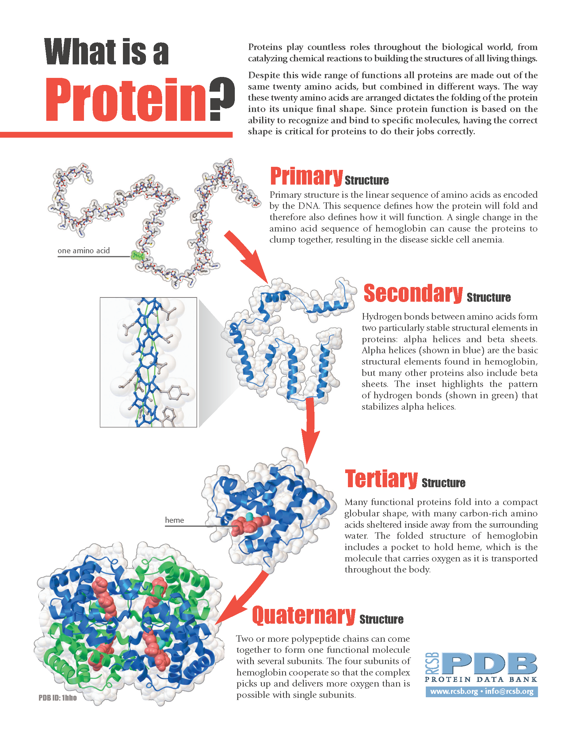
| Level | What it describes | Main idea to remember |
|---|---|---|
| Primary structure | The order of amino acids in a polypeptide chain | Sequence |
| Secondary structure | Local folding into shapes such as alpha helices and beta pleated sheets | Hydrogen bonds cause local folding |
| Tertiary structure | The overall three-dimensional shape of one polypeptide chain | R-group interactions cause further folding |
| Quaternary structure | The arrangement of two or more polypeptide subunits | Subunits join to make one functional protein |
Primary structure: the amino-acid sequence
The primary structure is not simply “amino acids” or “a protein chain.” It is the specific sequence of amino acids.
For example, these sequences contain the same three amino acids but are not the same primary structure:
Changing the order changes the positions of the R groups. That may alter which R groups can interact as folding occurs.
Secondary structure: local bends and coils
Parts of the chain may fold into regular shapes, especially:
- an alpha helix, which is a spiral or coil;
- a beta pleated sheet, which is a folded sheet-like arrangement.
These structures are held in place mainly by hydrogen bonds between parts of the polypeptide backbone.
At this level, the protein is not yet necessarily in its final functional shape. Think of secondary structure as local sections of the chain beginning to organise.
Tertiary structure: the final shape of one chain
The tertiary structure is the complete three-dimensional shape of a single polypeptide chain.
It results mainly from interactions between R groups. Important interactions include:
- hydrogen bonds;
- ionic attractions between oppositely charged R groups;
- attraction between hydrophobic R groups;
- disulfide bonds, which can form between certain sulfur-containing R groups.
In a watery cell environment, hydrophobic R groups tend to be folded towards the inside of the protein, away from water. Hydrophilic R groups tend to face outwards, where they can interact with water. This is one major reason the sequence influences the final shape.
Quaternary structure: several chains together
Some proteins consist of more than one polypeptide chain. Their quaternary structure is the way these subunits fit together.
Haemoglobin, the oxygen-carrying protein in red blood cells, has four polypeptide subunits. It has two alpha subunits and two beta subunits. The way these subunits associate is essential for haemoglobin’s function.
Not all proteins have quaternary structure. A protein made from one polypeptide chain has primary, secondary, and tertiary structure, but no quaternary structure.
Why shape controls function
A protein’s three-dimensional shape gives it particular surfaces, binding sites, and chemical properties. These determine what it can interact with.
For an enzyme, the shape of the active site allows a particular substrate to bind. If the active site changes shape, the substrate may no longer fit, so the enzyme cannot catalyse the reaction effectively.
For an antibody, a specifically shaped binding region allows it to attach to one particular antigen.
For a structural protein such as keratin, the arrangement of polypeptide chains produces a strong material suitable for hair, nails, or skin structures.
For a transport protein such as haemoglobin, the shape and arrangement of subunits allow oxygen to bind and be transported in the blood.
Protein structure and variety - Revise: Proteins - BBC
Read this BBC Bitesize revision page to reinforce the core link between amino-acid sequence, protein folding, shape, and biological function.
In the section “Protein structure and variety,” read the core explanation. Focus on the wording that links sequence, folding, three-dimensional shape, and interactions with other molecules. Then read the “Functions of proteins” table, using the examples of enzymes, hormones, antibodies, and structural proteins to see why different protein shapes are needed.
The “key and lock” comparison is useful, but use it carefully. It does not mean proteins are rigid metal objects. It means that the shape and chemical properties of a protein determine what it can bind to or act upon.
One substitution can matter: sickle-cell haemoglobin
A powerful example of sequence affecting function is sickle-cell disease.
In normal haemoglobin, the sixth amino acid in the beta chain is glutamic acid. In sickle-cell haemoglobin, that amino acid is replaced by valine.
This is a single amino-acid substitution, but it has major consequences:
- Primary structure changes: valine replaces glutamic acid in the beta chain.
- R-group properties change: glutamic acid has a charged, hydrophilic R group, while valine has a nonpolar, hydrophobic R group.
- Interactions between haemoglobin molecules change: the altered haemoglobin molecules can stick together, especially when oxygen concentration is low.
- Fibres form inside red blood cells: the haemoglobin aggregates into long fibres.
- Red blood cells become sickle-shaped and less flexible: they may block small blood vessels and reduce efficient oxygen transport.
The key point is not simply “a mutation causes sickle cell.” A higher-quality explanation follows the structure-function chain:
Replacing glutamic acid with valine changes the primary structure of the haemoglobin beta chain. Because the two amino acids have different R-group properties, the haemoglobin folds and interacts differently. Sickle-cell haemoglobin molecules can aggregate into fibres, distorting red blood cells and reducing effective oxygen transport.
Orders of protein structure: primary, secondary, tertiary ...
Read Khan Academy’s “Primary structure” section for the sickle-cell haemoglobin example, which shows how a small sequence change can cause a major change in protein behaviour.
In the “Primary structure” section, start at the paragraph beginning sequence and function. Then continue from the sentence beginning the sickle cell example. Focus on the causal chain from amino-acid substitution to altered haemoglobin behaviour and distorted red blood cells.
A single amino-acid substitution does not always cause a major change in function. Its effect depends on where it occurs and whether the replacement changes important interactions. However, the sickle-cell example shows why even one change can be significant.
Denaturation: when the sequence stays the same but shape changes
A protein’s sequence is important, but its environment is also important. Very high temperatures, extreme pH, or certain chemicals can disrupt the bonds and interactions holding the protein in its three-dimensional shape.
This is called denaturation.
During denaturation:
- the peptide bonds of the primary structure usually remain intact;
- secondary, tertiary, and sometimes quaternary structure are disrupted;
- the protein loses its specific shape;
- its function is reduced or lost.
For example, heating egg white denatures its proteins. The proteins unfold and form new interactions with each other, changing the egg white from clear liquid to a white solid.
This idea will become particularly important when you study enzymes: if temperature or pH changes an enzyme’s active site, the substrate may no longer fit.
Writing an assessment-quality explanation
For a question such as, “Explain how amino-acid sequence determines protein function,” avoid a one-step answer such as “the sequence determines function.” Include the mechanism.
Use this structure:
The amino-acid sequence is the protein’s primary structure. Different amino acids have different R groups, so the sequence determines which R groups occur and where they occur along the polypeptide. Interactions between these R groups cause the protein to fold into a specific three-dimensional shape. This shape determines what molecules the protein can bind to or act upon, so it determines the protein’s biological function.
For a question involving a mutation or changed amino acid, add:
A changed amino acid can have a different R group, altering interactions within or between protein molecules. This can change the protein’s shape and therefore its function.
Key takeaways
- Proteins are polypeptides made from amino acids joined by peptide bonds.
- The primary structure is the exact sequence of amino acids.
- Different amino acids have different R groups, with different chemical properties.
- R-group interactions cause the polypeptide to fold into a particular three-dimensional shape.
- A protein’s shape determines what it can bind to, transport, catalyse, or form; therefore, shape determines function.
- Haemoglobin in sickle-cell disease shows how one amino-acid substitution can alter protein interactions, cell shape, and body function.
- High temperature, extreme pH, or chemicals can denature proteins by disrupting their shape, even when the primary amino-acid sequence remains unchanged.
Next, you will move from protein structure to a practical written-test skill: identifying proteins and other nutrients using food-test procedures and their positive results.
Can't find a good explanation? Sign up and we'll make it for you
Sign up