
Hello! Welcome back to your course on Medical Instrumentation.
In our last lesson, we focused on the "how" of EEG analysis, learning to use spectral analysis techniques like the FFT to decompose a raw EEG signal into its fundamental frequency bands: Delta, Theta, Alpha, Beta, and Gamma.
Today, we transition from "how" to "what." We will explore the meaning behind these bands, directly addressing the learning outcome: Correlate specific EEG frequency band activity with cognitive and emotional states (e.g., arousal, attention, relaxation). This lesson is at the very heart of your work at Neuraease, as it bridges the gap between the signals your wearable devices collect and the human experiences you aim to understand.
A Deeper Dive into the Rhythms of the Brain
Last time, we created a basic table of the frequency bands and their associated states. Now, we'll explore the nuances of each one. Brain rhythms are dynamic; their meaning can depend on where they appear on the scalp and what a person is doing.
To get a comprehensive overview from an expert, we'll watch selected segments of a lecture from the Harvard Brain Science Initiative. This will provide a rich narrative context for each frequency band.
The "Brain Rhythms 101" lecture provides a fantastic, in-depth look at the major brain oscillations. We will watch specific clips to understand the cognitive function and underlying mechanisms of each band.
Watch the following segments. For each one, focus on the key cognitive states and functions associated with the rhythm, and note that these 'rules' are often generalizations. Delta (δ) Waves (1-4 Hz): Watch from 14:43 to 17:00. Note its role in deep sleep and as a non-specific marker of brain injury. Theta (θ) Waves (4-8 Hz): Watch from 20:26 to 23:20. Focus on its link to drowsiness, memory, and spatial navigation. Alpha (α) Waves (8-12 Hz): Watch from 25:54 to 29:30. Pay close attention to its classic presentation (eyes-closed, relaxed) and the concept of it being an 'inhibitory' or 'gating' mechanism. Beta (β) Waves (12-30 Hz): Watch from 31:39 to 34:15. Note its presence in normal wakefulness and its interesting role in motor control (suppression during movement). Gamma (γ) Waves (>30 Hz): Watch from 41:50 to 45:16. Understand its association with high-level processing, attention, and the 'binding problem.' Cross-Frequency Coupling: Watch from 45:16 to 47:00. This is an advanced but crucial concept: bands don't work in isolation; lower frequencies often modulate higher frequencies. Summary: Watch from 52:26 to 54:21 for a concise recap.
For a more structured, text-based reference that will be useful for exam preparation, the "Normal EEG Waveforms" article from StatPearls is an excellent resource. You can use it to solidify your understanding.
Normal EEG Waveforms - StatPearls
This clinical reference provides detailed descriptions of each EEG rhythm, including their typical locations, associated states, and pathological significance. It's a great resource to bookmark for future study.
Skim the sections on δ Rhythm, θ Rhythm, α Rhythm, β Rhythm, and γ Waves. You don't need to memorize every detail, but notice how the descriptions align with and expand upon the video lecture. Pay particular attention to the links made to cognitive processes like memory, attention, and decision-making.
Here's a refined summary table incorporating these new details, which you might find useful for quick reference.
| Band | Frequency | Key Cognitive & Emotional Correlates |
|---|---|---|
| Delta (δ) | 0.5 – 4 Hz | Low Arousal: Deep (slow-wave) sleep. Pathological if dominant during wakefulness (indicates brain injury). |
| Theta (θ) | 4 – 8 Hz | Drowsiness/Relaxation: Light sleep, deep meditation. Cognitive Control: Memory consolidation, frontal theta linked to processing conflict/effort. |
| Alpha (α) | 8 – 12 Hz | Relaxed Wakefulness: Dominant over the posterior scalp with eyes closed ("idling"). Suppressed by eye-opening and mental effort. Acts as a sensory "gate." |
| Beta (β) | 12 – 30 Hz | Active Wakefulness: Normal waking consciousness, active thinking, focus, alertness. Also involved in motor control. |
| Gamma (γ) | > 30 Hz | High-Level Processing: "Binding" sensory inputs into a coherent whole, peak focus, intense concentration, working memory. |
Modeling Emotion: The Valence-Arousal Space
Correlating EEG with "emotion" is complex because emotions themselves are complex. Are "anger" and "fear" fundamentally different, or do they share properties? Psychologists often simplify emotions into two core dimensions: Valence and Arousal.
- Valence: The pleasantness or unpleasantness of an emotion. Ranges from negative (e.g., sad, stressed) to positive (e.g., happy, elated).
- Arousal: The intensity or energy level of an emotion. Ranges from low (e.g., calm, bored) to high (e.g., excited, anxious).
This forms a two-dimensional map called the Arousal-Valence Space, where any emotional state can be plotted.
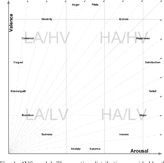
This model is incredibly useful in affective computing because it turns a subjective feeling into a quantifiable target. For your work at Neuraease, tracking a user's movement within this 2D space is more practical than trying to classify dozens of distinct emotions.
Let's see how this is used in a real research context.
Classification model of arousal and valence mental states by EEG
This research paper demonstrates a practical application of these concepts. It uses EEG signals to classify emotional states that have been defined using the Arousal-Valence model.
Read the following sections: Section II. EMOTIONS: Focus on the definitions of Arousal, Valence, and Dominance. Note how the authors create four classes (e.g., HA-HV for Happiness) based on this model. Table IV: Brain rhythms: This is a quick confirmation of the frequency bands we're studying. Section VI. CONCLUSIONS: Read the summary to understand the key finding: that a correlation exists between cortical electrical activity and moods, and that it can be classified computationally.
Connecting EEG Bands to Arousal and Valence
Now we can put everything together. How do the EEG bands we measure relate to the Arousal-Valence model? While this is an active area of research, some general principles have emerged:
-
Arousal Correlates with Frequency: In a very general sense, the overall frequency content of the EEG spectrum is linked to arousal.
- Low Arousal (calm, relaxed, drowsy) is characterized by lower-frequency activity dominating, particularly Alpha and Theta waves.
- High Arousal (alert, excited, stressed) is characterized by higher-frequency activity, particularly Beta and Gamma waves. A decrease in alpha power is also a strong indicator of rising arousal.
-
Valence is More Complex (Frontal Asymmetry): Valence doesn't have as simple a correlate, but one of the most studied markers is frontal alpha asymmetry.
- Positive Valence (happiness, joy) is often associated with relatively more alpha power in the right frontal lobe compared to the left (or, conversely, more left frontal activation).
- Negative Valence (sadness, disgust) is often associated with relatively more alpha power in the left frontal lobe compared to the right (or, conversely, more right frontal activation).
This is a powerful concept for your work. A simple ratio of frontal alpha power (Right - Left) / (Right + Left) can serve as a continuous measure of emotional valence.
Test your understanding!
A user of your Neuraease wearable is in a meeting. Your system detects the following simultaneous changes in their EEG:
- A significant drop in posterior Alpha power.
- A sharp increase in frontal Beta and Gamma power.
- A shift in frontal alpha asymmetry, indicating relatively more activity in the right frontal lobe compared to the left.
Based on the Arousal-Valence model, what cognitive/emotional state might this user be entering? Justify your answer based on each observation.
Show answer
The user is likely entering a state of High Arousal and Negative Valence, such as stress, anxiety, or frustration.
- High Arousal Justification: The drop in posterior Alpha power signifies a move away from a relaxed "idling" state. The increase in Beta and Gamma power indicates active, intense mental processing. Both are strong markers of high arousal.
- Negative Valence Justification: The shift towards relatively more right frontal activation (or less left frontal alpha power) is the classic indicator of negative valence.
This combination of features suggests the user is moving from a baseline state to one that is both mentally taxing and emotionally unpleasant—a prime candidate for a pre-meltdown warning.
Conclusion
In this lesson, you've moved beyond signal processing to signal interpretation. You now have a framework for translating the squiggly lines of an EEG into meaningful insights about a person's inner state.
Key Takeaways:
- Each primary EEG band (Delta, Theta, Alpha, Beta, Gamma) has a distinct correlation with cognitive functions and states of arousal.
- The Arousal-Valence model provides a practical, two-dimensional framework for quantifying complex emotional states.
- In general, arousal is reflected in the EEG's frequency content (higher frequency ≈ higher arousal).
- Valence is often studied through frontal alpha asymmetry, providing a potential marker for pleasant vs. unpleasant states.
Preview of the Next Lesson:
We've learned what the different rhythms mean and where they tend to show up. But how do we see this activity distributed across the entire head at a glance? In the next lesson, we will learn to perform quantitative EEG (qEEG) analysis to create topographical brain maps. This will allow us to visualize the spatial distribution of band power, turning abstract numbers into intuitive "heat maps" of brain activity.
Can't find a good explanation? Sign up and we'll make it for you
Sign up