
Hello! Welcome to the second lesson in our journey through the science of sleep.
In our previous lesson, we zoomed in to the microscopic level, exploring how individual brain cells—neurons—use chemical messengers called neurotransmitters to create a delicate balance between wakefulness and sleep. We introduced the idea of a "sleep-wake switch" controlled by competing excitatory (wake-promoting) and inhibitory (sleep-promoting) signals.
Today, we will zoom out to the macroscopic level. We will identify the specific command centers in the brain that house these neurons and orchestrate the complex symphony of sleep. Think of the last lesson as learning about the individual musicians and their instruments (neurons and neurotransmitters); this lesson is about meeting the conductors and the orchestra sections (brain regions).
By the end of this lesson, you will be able to identify the main brain regions involved in sleep regulation and understand their specific roles in helping you fall asleep, stay asleep, and wake up.
The Brain's Master Regulator: The Hypothalamus
Deep within your brain lies a small but incredibly powerful structure called the hypothalamus. Despite being only about the size of a peanut, it acts as a smart control and coordination center for many essential bodily functions, including hunger, thirst, body temperature, and, crucially, sleep.
To start, let's get a general overview of the key brain regions involved in sleep.
Brain Basics: Understanding Sleep
The following article from the National Institute of Neurological Disorders and Stroke (NINDS), titled 'Brain Basics: Understanding Sleep,' provides a great starting point. It clearly identifies the key brain structures and their functions.
Please read the section titled 'Anatomy of Sleep'. As you read, focus on creating a mental map of which parts of the brain are mentioned and their primary roles. We will explore each of these in more detail.
As the reading highlights, the hypothalamus contains several distinct groups of neurons that act as the core of the sleep-wake control system. Let's look at the most important ones.
The Sleep-Wake Switch: VLPO and Wake-Promoting Centers
Imagine a light switch. When you flip it one way, the light is on; when you flip it the other way, the light is off. A similar—though more complex—mechanism exists in your hypothalamus. Scientists call it the "flip-flop switch". It consists of two opposing groups of neurons that mutually inhibit each other.
-
Sleep-Promoting Center: The VLPO
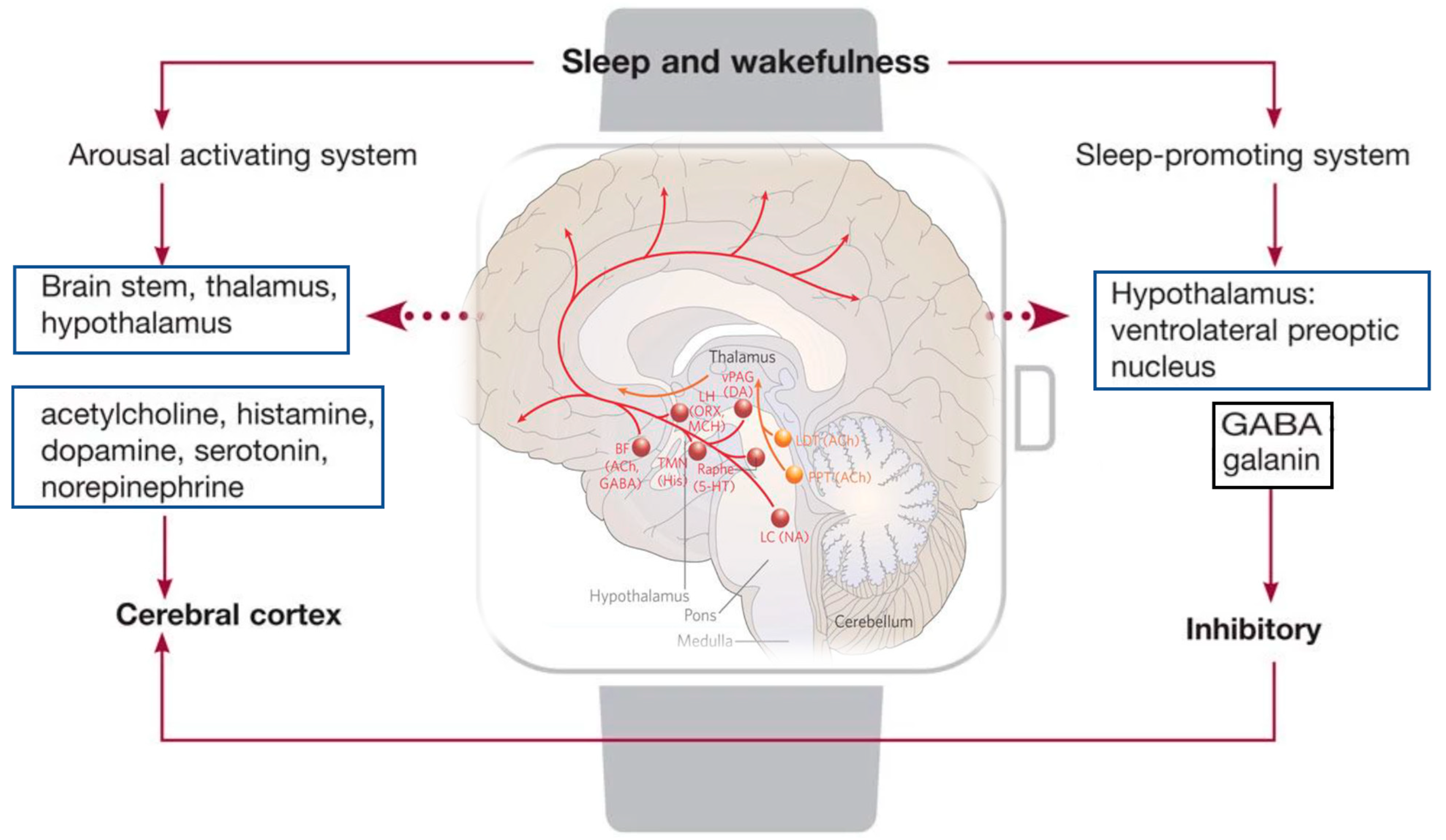
The ventrolateral preoptic nucleus (VLPO) is the brain's primary "sleep switch." When it's active, it releases the inhibitory neurotransmitter GABA (which we met in the last lesson) to dampen the activity of the brain's arousal centers. This action helps you fall asleep and stay asleep. -
Wake-Promoting Centers: The TMN and LH
On the other side of the switch are the arousal centers. These include the tuberomammillary nucleus (TMN), which releases the excitatory neurotransmitter histamine, and the lateral hypothalamus (LH), which releases orexin. When these areas are active, they promote wakefulness and alertness, and they also inhibit the VLPO, preventing you from falling asleep.
This mutual inhibition ensures that you are either mostly asleep or mostly awake, providing stability to each state.
The following short animation provides an excellent visual summary of this "switch" mechanism.
This clip from Alila Medical Media's 'Sleep Physiology' animation, which we briefly saw in the last lesson, perfectly illustrates the flip-flop switch in action. It shows how the VLPO and the wake-promoting regions oppose each other.
Please watch the segment from 3:48 to 5:15. Pay close attention to how the VLPO uses GABA to inhibit the wake-promoting regions (like the TMN) and how the SCN (which we'll discuss next) influences this switch.
This diagram shows the arousal and sleep-promoting systems in more detail. You can see the sleep-promoting VLPO at the top right, and the multiple wake-promoting centers in the brainstem and hypothalamus that it inhibits.
The Master Clock: The SCN
Also located in the hypothalamus is the suprachiasmatic nucleus (SCN). The SCN is your body's master biological clock. It receives information about light directly from your eyes and uses this information to align your internal rhythms with the 24-hour day-night cycle. The SCN helps orchestrate the timing of the flip-flop switch, sending out strong "wake up!" signals during the day to help the arousal centers overpower the sleep drive.
We will dedicate our entire next lesson to this fascinating structure and the circadian rhythms it controls.
Beyond the Hypothalamus: The Supporting Network
While the hypothalamus contains the central switch, it doesn't work in isolation. Other brain regions play critical supporting roles.
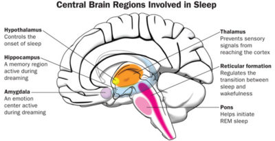
Let's review the functions of these other key players:
- The Brainstem (Pons, Medulla, Reticular Formation): This structure at the base of the brain connects the spinal cord to the rest of the brain. It's a crucial relay station and contains centers that help produce arousal. The brainstem is also fundamental for REM sleep. Specifically, the pons sends signals to temporarily paralyze your major muscles during dreams, preventing you from acting them out.
- The Thalamus: This region acts as the brain's sensory gatekeeper. During most of sleep (NREM sleep), the thalamus quiets down, blocking signals from your senses (sights, sounds, smells) from reaching the conscious part of your brain (the cortex). This is what allows you to tune out the external world. During REM sleep, however, the thalamus becomes active and sends the cortex the images and sounds that populate your dreams.
- The Basal Forebrain: Located near the front of the brain, this area also plays a role in promoting sleep. It is one of the areas where adenosine, the chemical that creates "sleep pressure," exerts its sleep-inducing effects.
- The Amygdala: Known as the brain's emotion center, the amygdala becomes highly active during REM sleep. This heightened activity is thought to be linked to the intense and often bizarre emotional content of our dreams.
Test your understanding!
A person is in a deep, dreamless sleep. An alarm clock goes off, but they don't wake up immediately. Which brain region is primarily responsible for blocking the sound of the alarm from reaching the person's conscious awareness?
Show answer
The thalamus. During deep non-REM sleep, the thalamus acts as a gate, preventing most sensory information (like the sound of the alarm) from being relayed to the cerebral cortex for conscious processing.
A Deeper Look: The Neural Circuits in Action
Now that we have identified the individual regions, let's watch a more detailed video that ties them together, explaining how these circuits control the different states of being awake, in non-REM (NREM) sleep, and in REM sleep.
The Neuroscience of Sleep: How The Brain Controls Sleep
The video 'The Neuroscience of Sleep' from the Sense of Mind channel offers a more in-depth look at the specific circuits that control wakefulness and sleep. It reinforces what we've learned and adds detail about REM sleep control.
Please watch the following segments: The Wakefulness Circuit (27:07 - 31:18): This section details the role of the lateral hypothalamus (LH) in keeping us awake by activating other brain regions. The NREM Sleep Circuit (36:25 - 41:43): This part focuses on the pre-optic area (where the VLPO is located) as the primary promoter of NREM sleep and its interaction with the wakefulness circuit. The REM Sleep Circuit (45:17 - 50:57): This explains that REM sleep control is centered in the brainstem (specifically the pons), distinct from the hypothalamic control of NREM sleep.
The video emphasizes an important point: while we can identify "control centers," the brain operates as a highly interconnected and dynamic network. Sleep is not governed by a single on/off button but by the shifting balance of activity across these multiple, overlapping regions.
Conclusion
In this lesson, we have mapped out the primary brain regions that form the complex network regulating sleep.
Key Takeaways:
- The hypothalamus is the central command center, containing the "flip-flop switch" for sleep and wakefulness.
- This switch involves a balance between the sleep-promoting VLPO (which uses GABA) and wake-promoting centers like the TMN (histamine) and LH (orexin).
- The SCN, also in the hypothalamus, acts as the master clock, timing the sleep-wake cycle.
- The brainstem is crucial for general arousal and is the specific control center for generating REM sleep and the associated muscle paralysis.
- The thalamus acts as a sensory gate, blocking external stimuli during sleep, but becomes active during dreaming to help generate dream content.
You now have a structural map of the sleeping brain, understanding not just what chemicals are involved, but where they act.
Preview of the Next Lesson:
We briefly introduced the suprachiasmatic nucleus (SCN) as the brain's master clock. In our next lesson, we will dive deep into its function. We will explore the concept of circadian rhythms—the 24-hour biological cycles that the SCN produces—and understand their profound role in governing our sleep-wake patterns.