
Hello! Welcome to the first lesson of our course.
Our journey together will explore the fascinating world of sleep, connecting modern scientific discoveries with the profound insights of Jain philosophy, particularly the teachings of Acharya Mahapragya on meditation and consciousness.
To begin this exploration, we must first build a solid foundation in the science of sleep. This initial module will focus on the biological machinery that governs our sleep-wake cycles. In this specific lesson, we will start with the most fundamental building blocks of the brain: the cells and the chemical signals they use to communicate.
By the end of this lesson, you will be able to describe the basic structure of a neuron, understand its function, and identify the key chemical messengers, or neurotransmitters, that play a crucial role in telling your brain when to be awake and when to fall asleep.
Let's begin by looking at the primary communicators of your nervous system.
The Brain's Messengers: An Introduction to Neurons
Your brain, and indeed your entire nervous system, is an intricate network of specialized cells called neurons. These are the fundamental units responsible for transmitting information, allowing you to think, feel, move, and perceive the world. To understand how sleep is regulated, we first need to understand how these cells are built and how they talk to each other.
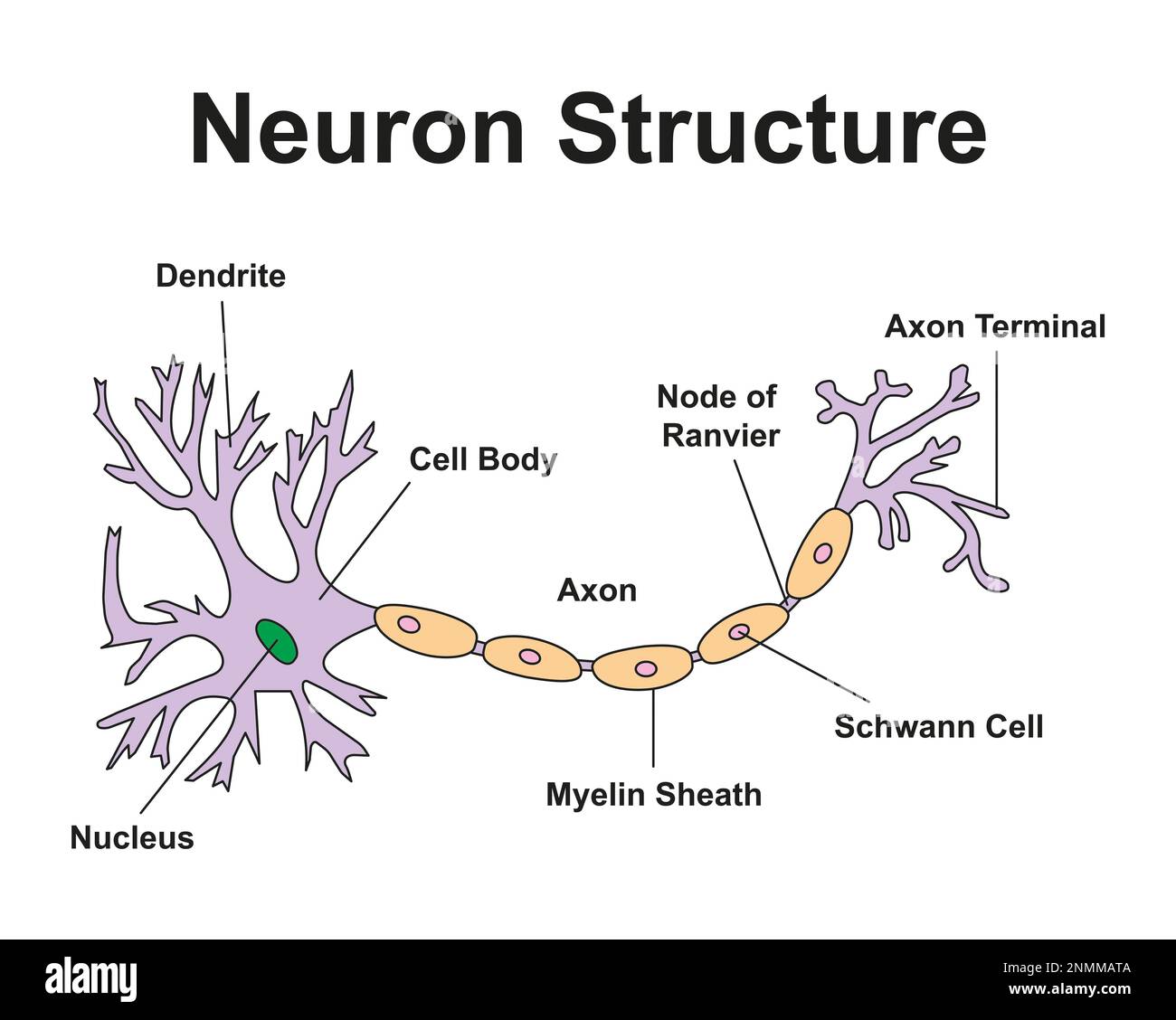
To get a dynamic understanding of how these parts work together, let's watch a video that breaks it down step-by-step.
What is a Neuron? Parts and Function
The following video, 'What is a Neuron? Parts and Function' from the Psych Explained channel, provides an excellent and clear introduction to the structure and function of a neuron. We'll watch it in segments to focus on key concepts.
Please watch the following parts of the video: Starting at 41 seconds, watch The Receiving End: Focus on the role of the dendrites as the receivers of chemical messages. Immediately after at 1 minute and 32 seconds, look at The Control Center: Understand the function of the soma (cell body) in integrating information. Skipping to 2 minutes and 32 seconds, check out The Transmission Cable: Learn about the axon, the long fiber that carries the signal away from the cell body. Finally, at 5 minutes and 55 seconds, observe The Sending End: Pay attention to the axon terminal and the concept of the synapse, the gap where communication with the next neuron happens.
As you saw in the video, a neuron's job is to pass messages along a chain. In essence:
- Receive: Dendrites collect incoming signals from other neurons.
- Integrate: The cell body (soma) processes these signals.
- Transmit: If the signal is strong enough, an electrical impulse (the action potential) travels down the axon.
- Communicate: At the axon terminal, the neuron releases chemical messengers to signal the next neuron in line.
This process of chemical communication is the key to everything the brain does, including falling asleep.
Test your understanding!
Imagine a message traveling from Neuron A to Neuron B. Which part of Neuron B receives the chemical signal sent by Neuron A's axon terminal?
Show answer
The dendrites of Neuron B would receive the signal. Dendrites are the specialized "receivers" of the neuron.
The Chemical Language of the Brain: Neurotransmitters
The chemical messengers that neurons use to communicate are called neurotransmitters. When the electrical signal reaches the end of a neuron (the axon terminal), it triggers the release of these neurotransmitters into the tiny gap between neurons, known as the synapse.
These molecules then travel across the synapse and bind to specific receptors on the dendrites of the next neuron, much like a key fitting into a lock.

This binding can have one of two general effects:
- Excitatory: It can encourage the receiving neuron to "fire" its own signal, passing the message along. Think of this as a "green light" or an accelerator.
- Inhibitory: It can discourage the receiving neuron from firing. Think of this as a "red light" or a brake pedal.
The regulation of sleep is a beautiful example of this interplay between excitatory and inhibitory signals. Different groups of neurons in your brain work to either promote wakefulness (excitatory) or promote sleep (inhibitory).
The Neurochemistry of Sleep and Wakefulness
Now, let's connect this foundational knowledge to our topic: sleep. The transition from wakefulness to sleep isn't like a simple on/off switch, but rather a carefully managed balance—a "flip-flop switch," as some scientists call it—controlled by competing groups of neurons.
To learn about the specific neurotransmitters involved, please read a short excerpt from an article by Harvard Medical School's Division of Sleep Medicine.
Science of Sleep: How is Sleep Regulated?
This reading, from 'Science of Sleep: How is Sleep Regulated?', will introduce you to the key wake-promoting and sleep-promoting chemicals and the brain areas that release them.
Please find and read the section titled 'Stable Wakefulness and Stable Sleep'. Below the heading, read about sleep-wake regulation. Focus on identifying the neurotransmitters mentioned (histamine and orexin) and their role in promoting arousal, as well as the brain area responsible for shutting down these arousal signals (the VLPO).
The reading highlights two key players in keeping you awake:
- Histamine: This is an excitatory neurotransmitter that promotes arousal. This is why antihistamine medications, which block histamine receptors, often cause drowsiness.
- Orexin (also called Hypocretin): This is another powerful wake-promoting neurotransmitter. A loss of the neurons that produce orexin leads to narcolepsy, a disorder characterized by overwhelming daytime sleepiness.
To promote sleep, other neurons, particularly in a brain region called the VLPO, release inhibitory neurotransmitters that "turn off" these arousal centers.
Let's broaden our view with another short reading that lists the main neurotransmitters involved.
Brain Basics: Understanding Sleep
This excerpt from the National Institute of Neurological Disorders and Stroke (NINDS) provides a concise list of the key chemical players in the sleep-wake cycle.
Please read the short paragraph under the heading 'Chemical signals to sleep' about the chemical signals. As you read, make a mental note of the neurotransmitters listed and their general role.
Combining what we've learned, we can categorize the main neurotransmitters involved in sleep and wakefulness:
| Role | Neurotransmitter | Primary Function |
|---|---|---|
| Wake-Promoting | Orexin/Hypocretin | Strongly promotes and sustains wakefulness. |
| Wake-Promoting | Histamine | Promotes arousal and alertness. |
| Wake-Promoting | Norepinephrine | Increases alertness, arousal, and attention. |
| Wake-Promoting | Acetylcholine | Active during wakefulness and REM sleep. |
| Sleep-Promoting | GABA | The main inhibitory neurotransmitter; it calms nerve activity. |
| Sleep-Promoting | Adenosine | Builds up during waking hours, creating "sleep pressure." |
Of these, GABA and Adenosine are particularly important for initiating and maintaining sleep.
- GABA (Gamma-aminobutyric acid) is the primary "brake" pedal of the brain. Sleep-promoting neurons in the VLPO use GABA to inhibit the wake-promoting regions.
- Adenosine is a byproduct of energy consumption in your cells. Its level steadily rises the longer you stay awake. This build-up increases the "drive to sleep" by inhibiting wake-promoting neurons. Caffeine works by blocking adenosine's action, which is how it keeps you alert.
The following animation provides a great summary of how these systems interact in a "sleep-wake switch."
This clip from 'Sleep Physiology' by Alila Medical Media visually summarizes the 'switch' mechanism we've been discussing, showing how brain regions use neurotransmitters to turn sleep on and off.
Starting a little before the four-minute mark, please watch the sleep-wake switch. Pay attention to how the VLPO (sleep-promoting region) uses GABA to inhibit the wake-promoting regions, like the TMN which releases histamine.
Conclusion
In this first lesson, we have laid the groundwork for understanding the biology of sleep by focusing on its smallest components.
Key Takeaways:
- The brain's basic communication cells are neurons, which receive, process, and transmit information.
- Neurons communicate with each other across a gap called a synapse using chemical messengers called neurotransmitters.
- These neurotransmitters can be excitatory (promoting action) or inhibitory (preventing action).
- The sleep-wake cycle is controlled by a balance between wake-promoting systems (using neurotransmitters like orexin and histamine) and sleep-promoting systems (using neurotransmitters like GABA and adenosine).
You now have a foundational vocabulary and conceptual model to understand the brain's activity during sleep.
Preview of the Next Lesson:
Now that we have seen how neurons and neurotransmitters function at a microscopic level, our next lesson will zoom out. We will explore the larger brain structures and regions that house these neuronal systems and orchestrate the complex process of sleep regulation.