
Welcome to the module on sensory balance control. Balance is not a single “talent” that some people have and others lack. It is an ongoing control process: your nervous system continually estimates where you are, notices movement or instability, and adjusts your eyes and muscles before a small sway becomes a step or a fall.
In this lesson, you will learn the three principal information sources behind that estimate: vision, the vestibular system in the inner ear, and the somatosensory system of skin, muscles, tendons, and joints. This provides the reasoning behind later supported balance drills: they are not simply tests of willpower, but ways to practise using and integrating information safely.
Balance is a continuously updated estimate
In practical terms, balance means keeping your body’s centre of mass over its base of support. When standing with both feet planted, your base of support is the area beneath and between your feet. If your body drifts too far toward an edge of that area, you need a correction: perhaps a subtle ankle adjustment, a shift at the hips, a reach for support, or a step.
The correction depends on a useful internal estimate of several things at once:
- Where is my body relative to the floor and gravity?
- Where are my feet, knees, trunk, and head relative to one another?
- Am I moving, or is the environment moving around me?
- How quickly is a change occurring?
- How much muscle activity is needed to remain stable?
Your brain does not obtain all this from one place. It combines three partially overlapping sensory reports:
- Visual information tells you about your body relative to the environment.
- Vestibular information tells you about head movement, acceleration, and orientation relative to gravity.
- Somatosensory information tells you about pressure, contact, muscle stretch, joint position, and body configuration.
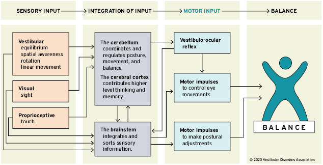
This is why balance can feel different in low light, on an uneven path, in a visually busy environment, or when getting up quickly. The same person has the same muscles in each situation, but the available information and the brain’s confidence in it have changed.
Vision: an external reference for orientation
Vision gives the nervous system a continuously changing map of the surroundings. Vertical doorframes, the floor, a counter, the edge of a pavement, and the apparent movement of nearby objects all help answer: “Where am I relative to the world?”
Both central and peripheral vision matter, but in different ways.
- Central vision is the focused, detailed vision you use to identify a curb, read a sign, or look at a fixed point.
- Peripheral vision is particularly useful for detecting broad movement around you and noticing sway relative to the room.
When you stand still and look at a stable feature in the room, visual input helps confirm that you are stable. When you walk, the changing visual scene provides information about direction and speed. This changing pattern is sometimes called optic flow: nearby objects seem to pass by faster than distant ones.
Vision is useful, but it is not always correct. Consider standing at a bus stop while a large bus beside you begins to move. For a moment, the movement in your visual field can create the impression that you are moving. Your feet and inner ear may disagree. The brain must resolve that mismatch.
A practical implication is that a visually complex setting can make balance more demanding even if the floor is perfectly safe. Later in the course, you may carefully vary visual conditions as a way to progress a task. For now, the sensible default during knee rehabilitation is to use a well-lit, visually stable space with reliable hand support available.
Somatosensory information: feeling contact and body position
The term somatosensory refers broadly to information from the body, including touch and pressure from the skin as well as position and movement information from muscles, tendons, and joints.
Proprioception is the part most relevant to body-position awareness. It is your sense of where body parts are and how they are moving, even without looking at them. You use it when you know that your knee is bent, feel that more weight is on one foot, or place a hand on a door handle without watching the whole movement.
Several types of information contribute:
- Pressure and touch receptors, especially in the soles of the feet, signal where and how firmly you contact the surface.
- Muscle receptors signal changes in muscle length and speed of stretch.
- Tendon receptors contribute information about muscle tension.
- Joint and skin receptors add information about joint angle, movement, and contact.
For standing balance, the feet and ankles are especially informative. A gentle forward lean increases pressure toward the front of the soles; a backward lean changes pressure toward the heels. Your nervous system uses those changing pressure patterns, along with muscle and joint signals, to estimate sway and make small corrections.
This is one reason a soft, sloped, slippery, or uneven surface feels different. It changes the quality and reliability of the information coming from the feet and ankles. It may also require different muscle responses. That does not mean you need to seek out unstable surfaces now. With your current restrictions, a firm, predictable floor and supported practice are the appropriate place to develop clear, repeatable sensory experience.
It is worth separating proprioception from interoception. Proprioception concerns body position and movement; interoception concerns internal states such as breathing, heart rate, temperature, hunger, fatigue, or nausea. Both can influence confident movement, but they are distinct systems. Interoception and effort regulation will be the focus of a later module.
The Human Balance System - Vestibular Disorders Association
Read “The Human Balance System” from the Vestibular Disorders Association for a clear account of the three sensory sources and how the brain combines them.
Start in “What is balance?” and “Sensory input.” Read the overview of sensory input to establish the three-source model. Then read the “Input from the eyes” subsection, especially the walking example, and notice how stable environmental features provide orientation. Next, read the full “Input from the muscles and joints” subsection. Focus on the explanation of proprioception, particularly the examples involving the soles of the feet, neck, and ankles. Continue with “Input from the vestibular system” and “Integration of sensory input.” Read the inner ear description, then the integration and sensory-conflict discussion. The bus example is a useful reminder that an odd sensation is not automatically an accurate account of what your body is doing.
The vestibular system: head movement and gravity
The vestibular system is located in the inner ear. It is not a “balance organ” in isolation; rather, it is a specialised source of information that is essential when your head moves or when visual and foot-based information are uncertain.
It has two main functional parts.
Semicircular canals: rotation
There are three semicircular canals on each side of the head, arranged in different planes. They detect angular acceleration: changes in the speed or direction of head rotation.
If you turn your head to look to the side, fluid in the relevant canal lags briefly because of inertia. This bends sensory structures and produces nerve signals. The signals tell the brain that your head is beginning to rotate and in which plane.
The canals are especially responsive to changes in rotation. This helps explain why spinning can cause dizziness: after you stop, fluid can continue moving briefly, so the vestibular signal says “movement” while visual and somatosensory information say “still.”
Otolith organs: gravity and linear acceleration
The utricle and saccule, known collectively as the otolith organs, provide information about head orientation relative to gravity and about linear acceleration or deceleration.
They contain tiny calcium-carbonate crystals embedded in a gel-like layer. Gravity and changes in motion shift that weighted layer relative to sensory hair cells. The resulting signal contributes to your sense of being upright, tilting, rising in a lift, or accelerating in a vehicle.
This information is especially important for body-position awareness because the brain needs to distinguish “my head is tilting” from “the visual world is moving” and from “my body is simply shifting weight over stable feet.”
The Vestibular System, Animation
Watch “The Vestibular System, Animation” by Alila Medical Media for a compact visual explanation of the inner-ear structures and their role in stable vision and posture.
Watch the overview and reflexes first. Focus on the distinction between conscious awareness of movement and rapid reflexive corrections that help stabilise gaze and posture. Then watch the inner ear anatomy. Notice the functional contrast: semicircular canals detect rotational head movement, while the otolith organs contribute information about gravity, head orientation, and linear acceleration. You do not need to memorise every anatomical label; aim to retain what each part detects.
One vestibular reflex is particularly relevant to everyday movement: the vestibulo-ocular reflex, or VOR. When you turn your head while looking at an object, your eyes automatically move in the opposite direction just enough to keep the image steady on the retina. This is why you can usually read a sign or keep your gaze on a person while walking.
In later practice, head movement may eventually be included as a balance variable. It should not be added casually during rehabilitation, because it changes the task substantially. For now, the important point is conceptual: stable vision during motion is an active achievement of the balance system, not merely something the eyes do on their own.
Integration: the brain decides what to rely on
The three sensory systems do not simply “vote,” with two sources outvoting the third. The nervous system assesses how reliable each source seems in the current context and gives it more or less influence.
On a firm floor in good light, vision and information from the feet may be highly reliable. On an uneven path at dusk, foot information may be noisier and vision less clear, so the brain may need to rely relatively more on vestibular cues and cautious movement. If a busy visual scene is misleading, stable pressure beneath the feet and vestibular signals can help correct the impression.
This flexible use of sensory information is often called sensory reweighting. It is one reason balance can improve with practice: practice teaches the nervous system which signals are useful in a particular task and how to produce an appropriately sized correction.
Several brain regions contribute:
- The brainstem rapidly processes sensory signals and supports automatic postural and eye-movement responses.
- The cerebellum helps calibrate timing, coordination, error correction, and learned movement patterns.
- The cerebral cortex contributes attention, planning, conscious perception, and remembered experience.
A dancer’s ability to remain poised through a turn or a gymnast’s ability to land accurately does not come from having one exceptional “balance sensor.” It reflects repeated calibration of these systems: detecting movement, predicting its consequences, correcting errors, and gradually making the response more automatic. The same learning principles apply at later adulthood, although the progression should be appropriately gradual and shaped by current physical restrictions.
A safe sensory observation
This is not a balance test and not a reason to reduce your hand support. It is simply a way to notice the three information streams during a movement already cleared by your physiotherapist.
For two or three minutes, in supportive footwear if that is how you usually do rehabilitation practice, stand at a stable kitchen counter or other approved support. Keep both feet planted and keep your gaze on a stable point ahead. Perform only the small, clinician-approved side-to-side or forward-back weight shifts already included in your programme. Do not pivot, twist the knee, close your eyes, or add head turns.
As you shift, quietly notice:
- Visual: the stable point ahead and any apparent movement of the room.
- Somatosensory: changing pressure under each foot, especially heel, forefoot, and outer or inner edge.
- Vestibular: the sense of head and body orientation relative to upright and gravity.
The goal is not to force a vivid sensation or to judge yourself. It is to connect language to experience. With repetition, you may become better able to describe what information you are using and when a movement begins to feel less certain.
Stop and return to a stable position if you experience pain, knee instability, a sudden increase in dizziness, visual disturbance, or a symptom that your rehabilitation plan identifies as a warning sign. New or persistent dizziness outside normal exertion warrants discussion with an appropriate clinician.
What this means for future balance practice
A well-designed balance drill changes the demands on one or more sensory systems while keeping the task safe enough for useful learning. For example, changing hand support, stance width, surface firmness, visual complexity, or head movement can alter the information available. But changing several at once makes it difficult to know why the task became harder and can exceed your current safety margin.
For your present stage, use this principle:
Keep the environment stable, maintain approved support, and use predictable movements while learning to notice sensory information.
Later lessons will use the same principle to progress or regress a task by changing one variable at a time. This is more useful than trying to imitate advanced dancers or gymnasts, whose performance reflects years of accumulated skill and tolerance for far more demanding conditions.
Key takeaways
Balance and body-position awareness depend on the integration of three main information sources:
- Vision provides an external reference for orientation and motion.
- Somatosensory and proprioceptive input provides information about contact, pressure, body configuration, and movement from the skin, muscles, tendons, and joints.
- Vestibular input provides information about head rotation, linear acceleration, and orientation relative to gravity.
The brainstem, cerebellum, and cerebral cortex continually combine these signals and organise responses in the eyes and postural muscles. When signals disagree, the resulting uncertainty or disorientation is understandable; it is not necessarily evidence that any single sense has “failed.”
Next, you will apply this model in supported weight shifts and stance variations, using one agreed movement-quality criterion to keep practice both safe and informative.
Can't find a good explanation? Sign up and we'll make it for you
Sign up