
Hello! Welcome to the next lesson in our journey through biomedical instrumentation.
In the last lesson, we focused on the somatic nervous system by designing a system to measure Electromyography (EMG). We saw how EMG captures the electrical signals from voluntary muscle contractions. Today, we shift our focus from voluntary control to the body's automatic, involuntary responses by exploring a key signal from the autonomic nervous system.
This lesson addresses the learning outcome: Explain the physiological basis of Electrodermal Activity (EDA) and its relation to sympathetic nervous system arousal. EDA is one of the most widely used signals for tracking stress, arousal, and emotional engagement, making it highly relevant to the work you do at Neuraease. We will uncover what EDA is, how the body produces it, and what its different components tell us.
1. What is Electrodermal Activity (EDA)?
At its core, EDA refers to the continuous variation in the electrical characteristics of the skin. While we previously measured the electrical activity of muscles, here we are measuring how the skin's ability to conduct electricity changes in response to psychological stimuli.
You may encounter several terms for this phenomenon:
- Galvanic Skin Response (GSR): An older term you will still see frequently.
- Skin Conductance (SC): This specifically refers to the most common method of measuring EDA.
- Electrodermal Activity (EDA): The modern, umbrella term that covers all related measurements.
The image below gives a simple but effective overview of the concept.
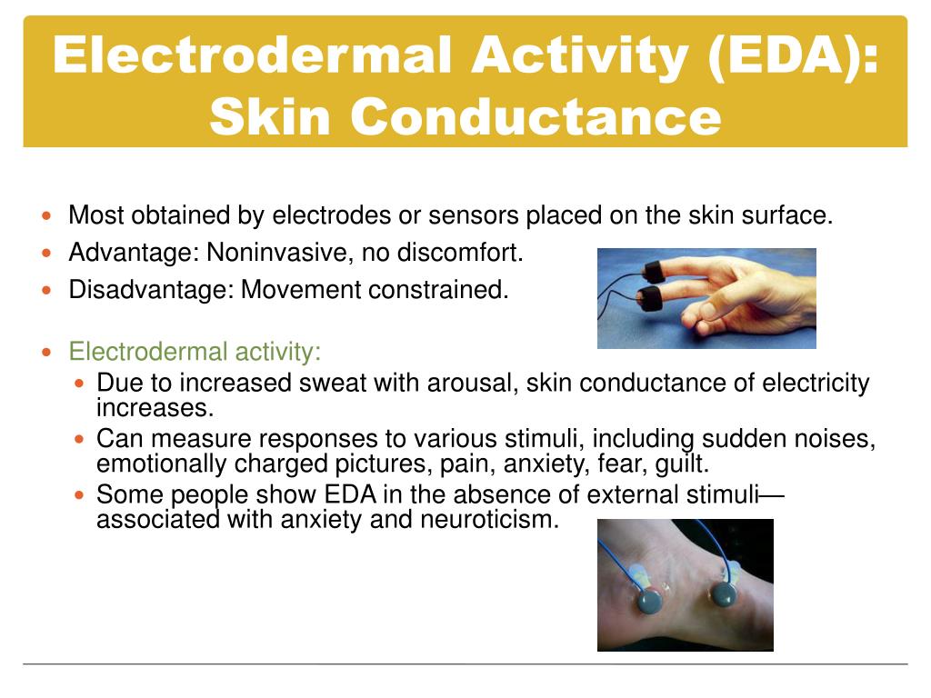
To get a more formal definition, let's turn to a short reading.
Electrodermal Response - an overview
The article 'Electrodermal Response - an overview' on ScienceDirect provides a clear and concise introduction to the topic.
Please read the section titled '1. Introduction to Electrodermal Response in Neuroscience'. This section defines the key terms (EDA, GSR, SCR) and introduces the two main components of the signal, which we will explore in detail shortly.
As the reading clarifies, EDA is fundamentally a measure of sweat gland activity, which in turn is a proxy for the activation of a specific part of our nervous system.
2. The Physiological Basis: The Sympathetic Nervous System and Sweat
To understand why EDA is such a powerful indicator of arousal, we need to look at the Autonomic Nervous System (ANS). The ANS controls our involuntary bodily functions like heart rate, digestion, and breathing. It has two main branches that often work in opposition:
- Parasympathetic Nervous System (PNS): The "rest and digest" system. It's dominant during calm, relaxed states.
- Sympathetic Nervous System (SNS): The "fight or flight" system. It prepares the body for action and is activated by stress, excitement, fear, and other emotionally salient events.
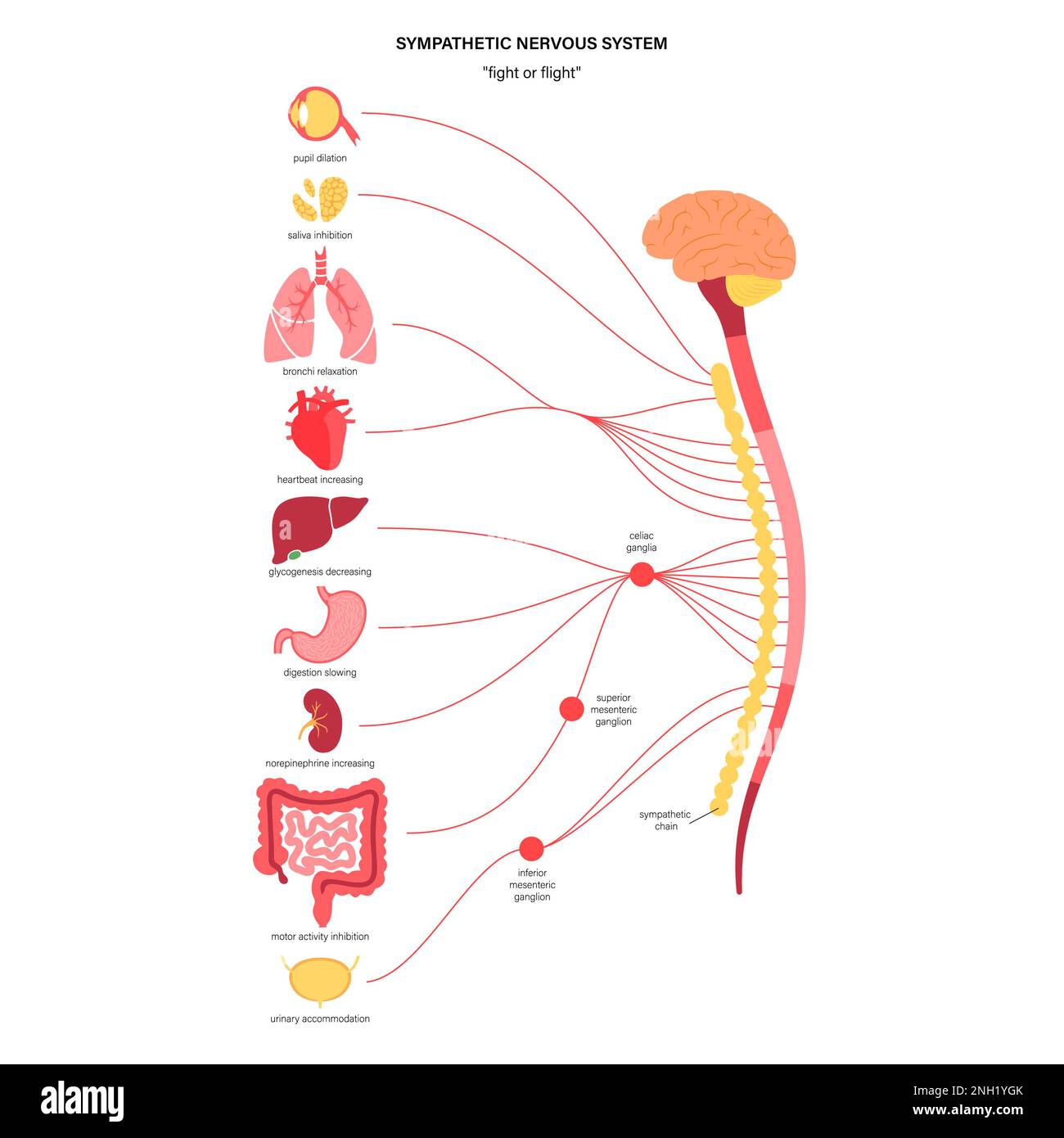
The key to EDA lies in a unique physiological fact: the eccrine sweat glands located on the palms of our hands and the soles of our feet are innervated exclusively by the Sympathetic Nervous System. They don't receive any input from the parasympathetic branch. This makes them a pure, unadulterated window into sympathetic arousal.
Here’s the chain of events:
- An emotionally relevant stimulus (e.g., a sudden noise, an anxious thought, a difficult task) triggers the SNS.
- The SNS sends nerve signals to the eccrine sweat glands. Unusually for the SNS, the neurotransmitter released at these glands is acetylcholine, not norepinephrine.
- The sweat glands secrete sweat, which is essentially a weak saline solution (salt water).
- This sweat rises up the sweat ducts, hydrating the dry outer layer of skin (the stratum corneum) and filling the ducts themselves.
- Since salt water is an excellent electrical conductor, this process dramatically lowers the electrical resistance of the skin.
- In electronics, conductance is the reciprocal of resistance (). Therefore, as skin resistance decreases, skin conductance increases.
This direct, simple link between sympathetic arousal and a measurable electrical property is what makes EDA so valuable for assessing emotional and cognitive states.
3. The Two Components of EDA: Tonic and Phasic
The EDA signal is not monolithic; it's composed of two distinct components that provide different kinds of information. Understanding this distinction is critical for practical applications.
Electrodermal Response - an overview
Let's revisit the ScienceDirect article to understand these two components.
Please re-read the second and last paragraphs of section '1. Introduction to Electrodermal Response in Neuroscience' and the summary paragraph in section '4. Applications of Electrodermal Response...'. Focus on the distinction between the 'phasic' component (SCR) and the 'tonic' component (SCL).
Tonic Activity: Skin Conductance Level (SCL)
The tonic component, known as the Skin Conductance Level (SCL), is the slow-changing, underlying baseline level of conductance.
- What it represents: General arousal, alertness, or stress level over longer periods (seconds to minutes).
- Signal characteristics: A slowly drifting signal. It tends to rise during periods of stress or high cognitive load and fall during relaxation.
- Analogy: Think of it as the overall water level in a reservoir. It changes slowly based on inflow and outflow.
- Application for Neuraease: Tracking a slow, steady rise in SCL over several minutes could be a powerful indicator of mounting stress or cognitive overload, a key precursor to a potential meltdown.
Phasic Activity: Skin Conductance Response (SCR)
The phasic component, known as the Skin Conductance Response (SCR), consists of rapid, transient increases in conductance.
- What it represents: A brief, specific response to a discrete stimulus—be it external (a loud noise) or internal (a sudden memory or thought).
- Signal characteristics: A distinct peak that rises 1-4 seconds after a stimulus and then slowly recovers to the baseline SCL.
- Analogy: These are the waves or ripples on the surface of the reservoir caused by a stone being dropped in.
- Application for Neuraease: Detecting a high frequency of SCRs (many small "jumps" in arousal) could signify a state of hyper-reactivity or sensory sensitivity, another important feature to track.
Test your understanding!
Imagine you are analyzing data from a wearable device for a user in a classroom. You observe two distinct patterns in their EDA data at different times.
- Pattern A: During a pop quiz, the baseline EDA level slowly and steadily climbs to double its initial value over 10 minutes.
- Pattern B: While listening to a lecture, the baseline is stable, but there is a sharp, distinct peak in the signal every time the teacher bangs a ruler on the desk to get the class's attention.
Which pattern corresponds to a change in SCL, and which corresponds to an SCR? What does each pattern tell you about the user's state?
Show answer
- Pattern A is a change in SCL (Tonic activity). The slow, sustained increase in the baseline reflects a general rise in physiological arousal and stress caused by the cognitive demand and pressure of the pop quiz.
- Pattern B shows multiple SCRs (Phasic activity). Each sharp peak is a direct, involuntary orienting response to a discrete, startling external stimulus (the ruler bang). The stable baseline (SCL) between the bangs suggests the user's general arousal level is not changing.
4. A Brief Look at Measurement
While a deep dive into the specific circuits for EDA is for another lesson, the basic measurement principle is straightforward and builds on your ECE foundation. It's an exosomatic method, meaning we apply a small external current.
The standard technique involves:
- Placing two electrodes on a site rich in eccrine glands, like the fingers or palm.
- Applying a very small, constant voltage (typically 0.5 V) across the electrodes.
- Measuring the resulting current that flows through the skin.
- According to Ohm's Law (), if the voltage () is constant, the measured current () is directly proportional to the skin's conductance (), since .
The resulting signal is measured in microsiemens (µS). The technical paper "Publication recommendations for electrodermal..." provides exhaustive detail on this DC constant-voltage method in section 1.2 if you're curious about the deeper specifics.
Conclusion
In this lesson, we've unraveled the physiological mechanism behind Electrodermal Activity, establishing it as a direct and reliable measure of sympathetic nervous system arousal. This makes it an invaluable tool for your work in assessing stress and emotional states.
Key Takeaways:
- EDA measures changes in skin conductance, which are caused by the activity of eccrine sweat glands.
- These specific sweat glands (on palms/soles) are controlled exclusively by the Sympathetic Nervous System (SNS), making EDA a pure index of "fight or flight" arousal.
- The signal has two components:
- Skin Conductance Level (SCL): The tonic, slow-changing baseline that reflects general arousal.
- Skin Conductance Response (SCR): The phasic, rapid peaks that reflect responses to specific stimuli.
- The mechanism is simple: SNS activation -> sweating -> lower skin resistance -> higher skin conductance.
Preview of the Next Lesson:
We've now looked at a pure marker of sympathetic activity. But what about the other side of the coin, the parasympathetic "rest and digest" system? In our next lesson, we will explore Heart Rate Variability (HRV). We'll learn how analyzing the tiny variations in time between heartbeats can give us profound insight into the balance between the sympathetic and parasympathetic systems, providing an even more complete picture of autonomic function and resilience.
Can't find a good explanation? Sign up and we'll make it for you
Sign up