
Welcome to your first lesson in Medical Instrumentation!
This course is designed to provide you with the engineering principles needed to measure physiological data, which aligns directly with your work at Neuraease. We'll balance the theoretical knowledge required for your university exams with the practical insights you can apply to developing wearable neurotechnology.
Today, we'll begin by exploring the fundamental question: Why is it so challenging to measure signals from the human body? This lesson addresses the learning outcome, "Describe the unique challenges of measuring signals from living systems (e.g., safety, artifacts, variability)." Understanding these core problems is the essential first step before we can design solutions.
Let's start with a high-level view of what a medical instrumentation system looks like.
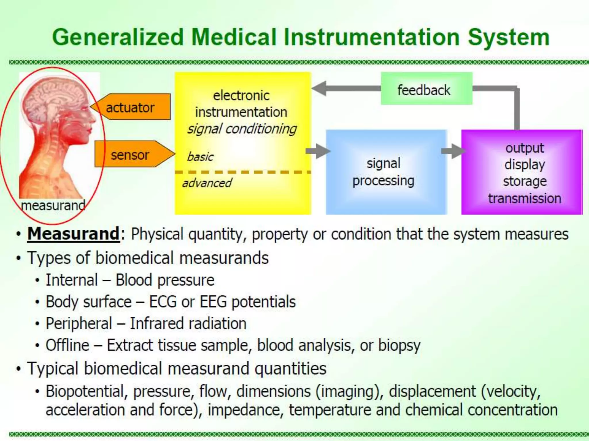
We will structure today's lesson around three major categories of these challenges:
- The Nature of the Source: Problems inherent to the living system itself.
- Artifacts and Noise: Unwanted signals that contaminate our measurements.
- Patient Safety: The paramount concern of ensuring no harm is done.
1. The Living System as a "Black Box"
Unlike a manufactured electronic circuit where you can probe any point and expect a predictable output, the human body is far more complex and delicate.
To get a comprehensive overview of these inherent difficulties, please start by reading the following article. It provides a solid foundation for the topics we'll discuss.
Key factors that affect or limit Biomedical Measurements
This article, 'Key factors that affect or limit Biomedical Measurements' from biomedicalinstrumentationsystems.com, clearly outlines the main challenges encountered when measuring signals from living systems.
Please read the entire article. Pay close attention to the sections titled 'Low measurement ranges', 'Inaccessibility of measurement variables', 'Hardly deterministic measurements', and 'Instruments operational constraints'.
As the article highlights, we face several fundamental problems:
- Inaccessibility: We often can't place a sensor at the ideal location. For example, measuring cardiac output directly would require an invasive procedure that isn't feasible for routine monitoring. We are forced to use indirect measurements from the body's surface, which brings its own set of complications.
- Variability: Biological signals are not deterministic. The same person's EEG can vary significantly based on their mental state, time of day, or even what they ate. Furthermore, signals vary widely between individuals. This natural variability is a major hurdle for systems like yours at Neuraease, which aim to identify consistent patterns related to specific states like an impending meltdown.
- Low Signal Strength: The physiological signals we're interested in are often incredibly faint. For instance, EEG signals measured on the scalp are in the range of microvolts ( V). These tiny signals are easily swamped by larger, unwanted electrical noise, which is why we'll spend a lot of time on amplification and filtering in later lessons.
2. Artifacts: The Unwanted Guests in Your Data
The tiny signals we want to measure are constantly competing with artifacts—unwanted signals that can corrupt or completely obscure the data. These are a primary enemy in biomedical instrumentation.
An artifact can come from the environment, the patient's own body, or the equipment itself. Let's watch a video that provides excellent, practical demonstrations of some of the most common artifacts in the context of an ECG recording. The principles are directly applicable to EEG, EMG, and other biopotential measurements.
ECG: common artefacts and how to avoid them
The video 'ECG: common artefacts and how to avoid them' from BPM biosignals will show you what artifacts look like in a real signal and how they are generated. This will make the concept much more tangible.
Watch the following segments: Mismatched Electrodes & Powerline Noise (00:51 - 02:40): Observe how a dried-out electrode allows 50 Hz powerline noise to dominate the signal. Muscle (EMG) Artifacts (02:40 - 04:30): See how simple arm movement generates large artifacts and how repositioning the electrode can mitigate this. Cable Motion Artifacts (05:25 - 06:56): Notice how even small movements of the electrode cable can introduce noise.
This video clearly demonstrated three key types of artifacts:
- Powerline Interference: The 50 Hz or 60 Hz hum from electrical wiring is everywhere. As the video showed, while differential amplifiers (which we will study in detail) are designed to reject this, their effectiveness depends on a balanced setup. Poor electrode contact unbalances the system and lets the noise in.
- Physiological Artifacts: Signals from other parts of the body can interfere. The most common example for brain-computer interfaces is contamination from muscle activity (EMG) during movement, or from eye blinks (EOG). For a wearable device, distinguishing a neural signal from the EMG of a tense jaw muscle is a critical challenge.
- Motion Artifacts: As shown with the cables, physical movement can create noise. This happens at the electrode-skin interface. The skin and electrode gel form a complex electrochemical junction. Movement changes the properties of this junction, generating spurious potentials that have nothing to do with the physiological signal of interest.
Test your understanding!
Imagine your startup, Neuraease, is testing a wearable headband to monitor EEG for signs of stress. A user reports that the device gives false alarms whenever they are chewing food. Based on what you've just learned, what is the most likely cause of these false alarms, and what type of artifact is it?
Show answer
The most likely cause is a physiological artifact specifically, Electromyographic (EMG) artifact. The chewing motion involves strong muscle contractions in the jaw (e.g., from the masseter and temporalis muscles). The electrical signals from these muscles are much larger than the underlying EEG signals from the brain and are likely being misinterpreted by the system as a neurological sign of stress.
3. Above All, Do No Harm: Patient Safety
When we attach an electronic device to a person, we create a potential pathway for electric current to enter the body. Ensuring this doesn't happen is the most important design constraint in medical instrumentation.
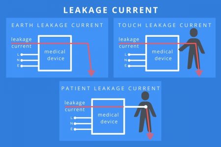
The danger comes from leakage current—small, unintended currents that can flow from a device to the ground, potentially through the patient. While these currents are often harmlessly small, a patient connected to a medical device can be uniquely vulnerable, especially if they have broken skin or internal connections (a condition known as being "electrically susceptible").
To standardize safety, medical devices are classified based on their level of patient protection. Let's watch a video that introduces these crucial safety concepts and classifications.
Electrical Safety Of Medical Equipment's | Biomedical Engineers TV |
This video, 'Electrical Safety Of Medical Equipment's' from Biomedical Engineers TV, covers the essential terminology and standards, including equipment classifications and leakage currents.
Please watch the following two clips: Equipment and Applied Part Classification (01:32 - 02:37): Focus on the definitions of Class 1/2 equipment and Type B, BF, and CF applied parts. Leakage Current Definitions (02:37 - 05:20): Listen for the definitions of earth leakage, enclosure (touch) leakage, and patient leakage current. Don't worry about memorizing every detail; focus on the general concept of what each one represents.
To summarize the key safety concepts:
- Applied Part: The part of the instrument that is in physical contact with the patient (e.g., an electrode).
- Classifications:
- Type B (Body): The least stringent; typically for devices not intended to be in direct contact with the patient.
- Type BF (Body Floating): The most common for surface-contact devices like ECG and EEG electrodes. The "F" stands for floating, meaning the applied part is electrically isolated from the rest of the device and earth ground, providing a crucial layer of protection. Your Neuraease device would need to meet Type BF standards.
- Type CF (Cardiac Floating): The most stringent classification, used for devices that will come in direct contact with the heart (e.g., internal pacemaker leads).
The video discussed different leakage currents. This diagram provides a helpful visual summary of what they are.
Finally, beyond electrical safety, we must also consider energy safety. Instruments like X-ray machines and diagnostic ultrasound actively transmit energy into the body. A key challenge is ensuring that this energy is kept at levels low enough to provide diagnostic information without causing tissue damage.
Conclusion
In this introductory lesson, we've seen that measuring signals from a living person is fundamentally different from measuring signals in a typical engineering system. The challenges are numerous and require careful consideration in every aspect of instrument design.
Key Takeaways:
- Inherent Challenges: Biological signals are often inaccessible, highly variable, and have very low amplitudes, making them difficult to isolate and interpret.
- Artifacts are Everywhere: Measurements are easily corrupted by electrical noise from the environment (powerlines), other physiological processes (EMG, EOG), and motion at the sensor interface.
- Safety is Paramount: Medical instruments must be designed to prevent hazardous electrical currents from passing through the patient. Safety classifications (like Type BF) and minimizing leakage currents are critical design requirements.
These challenges dictate the design of every biomedical instrument. They are the "why" behind the specific amplifier circuits, filtering techniques, and safety features we will be studying throughout this course.
Preview of the Next Lesson:
Now that we have an appreciation for the difficulties, our next step is to characterize the signals themselves. In Lesson 2, we will focus on the learning outcome: "Define key characteristics of biomedical signals, including typical amplitude and frequency ranges." We'll put numbers to concepts like "low signal strength" and start building a toolkit for how to capture them.
Can't find a good explanation? Sign up and we'll make it for you
Sign up