
Hello! Welcome to your next lesson.
In our last session, we explored how diagnostic ultrasound uses high-frequency sound waves to create images of soft tissues. We learned about the pulse-echo principle, acoustic impedance, and the crucial trade-off between imaging depth and resolution.
Today, we shift from imaging to functional testing as we examine the respiratory system. This lesson directly addresses the learning outcome: Explain the principles of spirometry for measuring lung volumes and capacities.
Spirometry is the most common pulmonary function test, providing essential data on how well the lungs are working. We will cover the key physiological metrics—lung volumes and capacities—and then dive into the engineering principles behind the instruments that measure them.
While this topic may seem distant from your startup's focus on neurotechnology, there are interesting links. Respiratory patterns are tightly coupled with the autonomic nervous system and emotional states. For example, changes in breathing can be a key indicator of stress or anxiety, so understanding the fundamentals of lung function provides a more complete picture of human physiology.
1. Defining Lung Volumes and Capacities
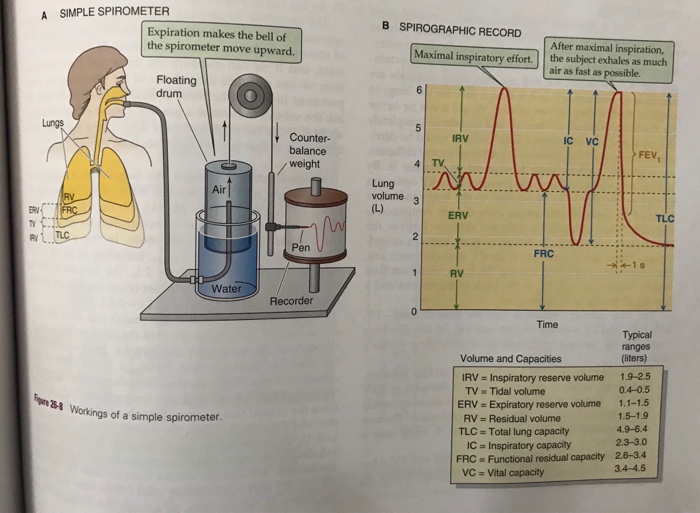
Before we can understand the instrument, we must first define what it measures. The air in your lungs can be divided into several distinct volumes. Combinations of these volumes are called capacities. These are typically visualized on a spirogram, which is a graph of lung volume versus time.
To get a clear understanding of these terms, let's watch a video that explains them with excellent visual aids and analogies.
Lung Volumes & Capacities : Foundations of Pulmonary Function Testing -Physiology USMLE Step 1
This video from Dr. G Bhanu Prakash provides a clear, step-by-step explanation of the four primary lung volumes and the four key lung capacities derived from them.
Please watch the video in segments, focusing on the following definitions: Lung Volumes (00:00 - 06:31): Pay attention to the definitions and visual representations of: Tidal Volume (TV) Inspiratory Reserve Volume (IRV) Expiratory Reserve Volume (ERV) Residual Volume (RV) Lung Capacities (07:20 - 15:18): Understand how capacities are combinations of the volumes you just learned: Inspiratory Capacity (IC) Vital Capacity (VC) Functional Residual Capacity (FRC) Total Lung Capacity (TLC)
Here is a summary of those key definitions:
Lung Volumes
- Tidal Volume (TV): The volume of air inhaled or exhaled during normal, quiet breathing.
- Inspiratory Reserve Volume (IRV): The maximum extra volume of air that can be forcefully inhaled after a normal tidal inhalation.
- Expiratory Reserve Volume (ERV): The maximum extra volume of air that can be forcefully exhaled after a normal tidal exhalation.
- Residual Volume (RV): The volume of air remaining in the lungs even after a maximal, forceful exhalation. This volume cannot be exhaled.
Lung Capacities
- Inspiratory Capacity (IC): The maximum volume of air that can be inhaled starting from the end of a normal exhalation.
- Functional Residual Capacity (FRC): The volume of air remaining in the lungs at the end of a normal, passive exhalation.
- Vital Capacity (VC): The maximum volume of air that can be exhaled after a maximal inhalation. This represents the total "usable" volume of the lungs.
- Total Lung Capacity (TLC): The total volume of air the lungs can hold.
What Spirometry Can and Cannot Measure
A crucial point from the video is that a spirometer only measures the air that moves in and out of the lungs. It cannot measure the air that remains trapped inside.
This means that Residual Volume (RV) cannot be measured by standard spirometry. Consequently, any capacity that includes RV in its calculation—namely, Functional Residual Capacity (FRC) and Total Lung Capacity (TLC)—also cannot be determined by spirometry alone. These values must be measured using other techniques like body plethysmography or gas dilution methods.
Test your understanding!
A patient's spirogram shows the following values:
- Tidal Volume (TV) = 500 mL
- Inspiratory Reserve Volume (IRV) = 3000 mL
- Expiratory Reserve Volume (ERV) = 1100 mL
- Calculate the patient's Vital Capacity (VC).
- Which of the following cannot be calculated from this information alone: Inspiratory Capacity (IC), Total Lung Capacity (TLC), or Vital Capacity (VC)?
Show answer
- Vital Capacity (VC) is the sum of IRV, TV, and ERV.
- Total Lung Capacity (TLC) cannot be calculated. To find TLC, you need to know the Residual Volume (RV), which cannot be measured by a standard spirometer.
2. Spirometer Instrumentation Principles
Now let's look at the engineering behind the instrument itself. Spirometers can be broadly categorized into two types: volume-sensing and flow-sensing.
Volume-Sensing Spirometers
The classic spirometer, as shown in Part A of the image Spirometer and Spirographic Record, is a volume-sensing device. It uses a bell or piston that is displaced by the patient's breath, directly measuring the volume of air moved. While highly accurate, these devices are bulky and less common in modern clinical practice.
Flow-Sensing Spirometers (Pneumotachographs)
Most modern spirometers are pneumotachographs, which measure airflow (). The volume () is then calculated by integrating the flow signal over time:
This approach will be familiar from your ECE background, where integrating current over time gives total charge.
The core principle of a pneumotachograph is analogous to Ohm's Law. It works by measuring the pressure drop () across a fixed resistance as air flows through it.

To get a better sense of the different engineering solutions used in modern spirometers, let's explore a few of the most common technologies.
The Different Types of Spirometer
The following resource from Vitalograph, a manufacturer of respiratory diagnostic devices, describes the working principles of several types of spirometer technologies. This will give you insight into the practical engineering behind these instruments.
Please read the sections on the following technologies. For each one, focus on the physical principle it uses to measure flow or volume: Fleisch Pneumotachograph: Found under its own heading. Note how it uses capillary tubes to create laminar flow and measures pressure drop. Stator-Rotor: Found under its own heading. Understand how it uses a spinning rotor and a light beam to measure flow. Rolling Seal: Found under its own heading. This is a modern, high-accuracy volume-sensing device. Note how it differs from the flow-sensing types. (Optional) Ultrasonic: Found under its own heading. See how it uses the Doppler effect on sound waves to measure flow.
As you can see, engineers have developed multiple ways to solve the problem of measuring airflow, each with its own trade-offs in terms of accuracy, cost, robustness, and hygiene. The pneumotachograph, particularly the Fleisch or Lilly (screen) type, remains a gold standard in clinical settings due to its linearity and accuracy.
Conclusion
In this lesson, we have explored the principles of spirometry, a cornerstone of respiratory diagnostics. We covered both the physiological measurements and the engineering behind the devices that acquire them.
Key Takeaways:
- Spirometry measures lung function by recording the movement of air during specific breathing maneuvers.
- The four primary lung volumes are Tidal Volume (TV), Inspiratory Reserve Volume (IRV), Expiratory Reserve Volume (ERV), and Residual Volume (RV).
- Lung capacities (IC, FRC, VC, TLC) are combinations of these volumes.
- Standard spirometry cannot measure RV, and therefore cannot determine FRC or TLC.
- Modern spirometers are often flow-sensing pneumotachographs that measure the pressure drop across a fixed resistance. Volume is then calculated by integrating the flow rate over time.
Preview of the Next Lesson:
We will continue our focus on respiratory diagnostics by looking at a ubiquitous clinical tool: the pulse oximeter. While spirometry assesses the mechanical function of the lungs (airflow and volumes), our next lesson will explain how we measure the physiological outcome of breathing: the oxygen saturation of the blood (SpO2).
Can't find a good explanation? Sign up and we'll make it for you
Sign up