
Hello! Welcome to the next step in our journey into EEG.
In the last lesson, we focused on the hardware, designing an acquisition system with high-quality amplifiers and filters to capture the cleanest possible signal. We engineered our system to fight noise with high input impedance, excellent common-mode rejection, and a Driven Right Leg circuit. However, in the real world, especially with wearable devices, some unwanted signals, or 'artifacts', will inevitably sneak past our hardware defenses.
This lesson directly addresses this challenge, fulfilling the learning outcome: Identify and apply techniques to mitigate common EEG artifacts (e.g., EOG, EMG, 60Hz noise).
We will shift our focus from hardware to procedural and software-based solutions. For your university exams, this is a frequent topic, testing your ability to ensure data integrity. For your work at Neuraease, this is arguably one of the most critical steps. The performance of any machine learning model you build to predict meltdowns is fundamentally dependent on its ability to distinguish true neural signals from contamination caused by a simple eye blink or muscle clench.
1. The Artifact Menagerie: What Are We Fighting?
First, let's formally define what we're up against.
EEG Artifacts: Types, Detection, and Removal Techniques ...
The article 'EEG Artifacts: Types, Detection, and Removal Techniques' from Bitbrain provides an excellent introduction. It defines what an EEG artifact is and gives a high-level categorization.
Please read the first three sections: 'What is an EEG artifact?', the 'Quick FAQ', and the introductory part of 'Types of EEG artifacts'. This will set the stage by defining the problem and its importance.
As the article explains, an EEG artifact is any electrical potential recorded by the EEG that does not originate from the brain's cortical activity. These artifacts are a major challenge because their amplitude can be 10 to 100 times larger than the actual EEG signal we want to measure.
Artifacts are broadly divided into two categories:
- Physiological Artifacts: Originating from the subject's own body (e.g., eye movements, muscle activity, heartbeat).
- Non-physiological (Technical) Artifacts: Originating from the environment or the equipment itself (e.g., 60Hz powerline noise, electrode movement).
To become proficient at cleaning EEG data, you first need to be an expert at visually identifying these intruders.
6 EEG Artifacts You Must Know in 6 Minutes
The video '6 EEG Artifacts You Must Know in 6 Minutes' by Jared Beckwith offers a quick and practical visual tour of the most common culprits. It's a great way to start building your visual recognition skills.
Watch the entire video (about 6 minutes). Pay close attention to the distinct shapes and characteristics of each artifact he presents: muscle (EMG), chewing, eye blinks (EOG), eye movements, cardiac (ECG), and 60Hz noise.
Now that you have a visual sense of these artifacts, let's organize them more formally.
2. A Field Guide to Common EEG Artifacts
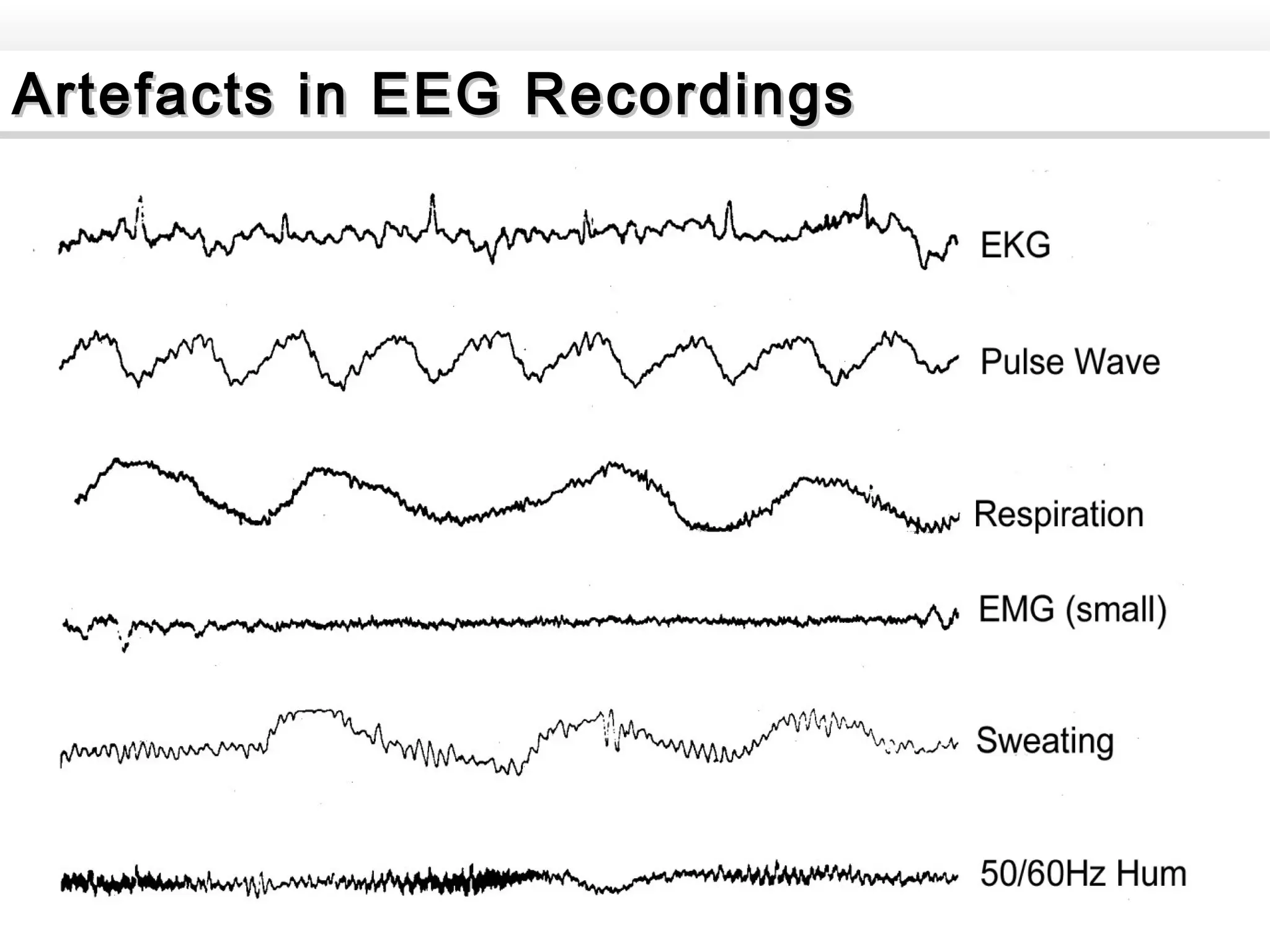
The following table, compiled from the Bitbrain article and the Mentalab resource "How To Reduce Noise In EEG Recordings", provides a more detailed reference.
| Artifact Type | Source | Typical Appearance & Location | Frequency Content |
|---|---|---|---|
| Eye Blinks (EOG) | Eyelid movement over the cornea-retinal dipole | Large (>100 µV), sharp, positive deflections. Strongest in frontal channels (Fp1, Fp2). | Low frequency (0.5 - 3 Hz), overlaps with Delta band. |
| Eye Movements (EOG) | Rotation of the eyeball dipole | Slower, rolling waves. Lateral movements cause out-of-phase signals in F7/F8. | Low frequency (0.5 - 3 Hz), overlaps with Delta band. |
| Muscle Activity (EMG) | Muscle contractions (jaw, neck, face) | High-frequency, spiky, "fuzzy" appearance. Can be brief bursts or sustained contamination. | Broadband, but primarily high frequency (> 20 Hz). |
| Cardiac (ECG/Pulse) | Heart's electrical activity or blood vessel pulse | Rhythmic, sharp spikes synchronized with the heart rate. | Can overlap with many EEG bands. |
| 60/50 Hz Powerline Noise | Electromagnetic interference from AC power | Persistent, high-frequency, sinusoidal "hum" across multiple channels. | A sharp peak at exactly 60 Hz (or 50 Hz). |
| Electrode 'Pop' | Sudden change in electrode impedance | Abrupt, large, transient spike or shift, often affecting only a single channel. | Broadband, transient. |
| Body/Head Movement | Physical movement causing electrode displacement | Slow, large, drifting waves that can saturate the amplifier. | Very low frequency (< 1 Hz). |
Test your understanding!
You are reviewing EEG data from a wearable sensor for Neuraease. You notice a rhythmic, sharp-peaked waveform occurring about once per second across several channels, but it's most prominent on the left side, near the T7 electrode. The participant was sitting still and was not blinking. What is the most likely artifact, and why?
Show answer
The most likely artifact is the cardiac artifact (ECG). The rhythmic occurrence at ~60 beats per minute (once per second) is the key clue. Its prominence can vary depending on electrode placement relative to major blood vessels. While a pulse artifact is possible, the "sharp-peaked" description is more characteristic of the QRS complex from the ECG signal itself bleeding into the EEG recording.
3. Mitigation Strategies: Before, During, and After Recording
Effective artifact removal isn't just a post-processing step; it's a comprehensive strategy. We can group our techniques into three phases.
How To Reduce Noise In EEG Recordings [11 Solutions]
The article 'How To Reduce Noise In EEG Recordings' provides a fantastic, practical framework for this. It outlines steps you can take before, during, and after recording.
Read the sections 'Before EEG recording' and 'During EEG recording'. These short sections offer simple, proactive measures that can dramatically improve data quality from the outset.
-
Before Recording (Proactive Mitigation):
- Participant Instructions: Instruct the participant to relax their jaw, minimize swallowing, and avoid excessive blinking, especially during critical parts of the recording.
- Environment Control: Use a room with minimal electrical equipment. Turn off fluorescent lights and computer monitors if possible. In a clinical setting, a Faraday cage might be used. For a wearable, this isn't possible, making post-processing even more critical.
-
During Recording (Procedural Mitigation):
- Good Electrode Contact: This is paramount. Ensure skin is properly prepped and electrode impedances are low and stable (as we discussed in a previous lesson).
- Secure Cables: Prevent cable movement, which can cause large artifacts. Taping down cables or using systems with integrated, short cables is effective. This is a major design consideration for wearables.
-
After Recording (Post-Processing):
- This is where we use software and algorithms to clean up the data that has been recorded. Even with the best preparation, this step is always necessary. We will focus on these techniques for the rest of the lesson.
4. Post-Processing Techniques for Artifact Removal
There are several computational approaches to cleaning EEG data, ranging from simple to highly advanced.
EEG Artifacts: Types, Detection, and Removal Techniques ...
Let's return to the Bitbrain article, which gives a great overview of the main data-driven filtering techniques.
Read the sections 'EEG Artifact Filtering Techniques (Data-Driven Approaches)' and 'EEG Artifact Filtering Techniques: Online vs. Offline Approaches'. This will introduce you to the main concepts of Rejection, Filtering, Blind Source Separation (BSS), and Source Decomposition.
Technique 1: Filtering and Rejection
- Filtering: This is the most straightforward approach. You'll recall from our hardware lesson that we used filters to define our signal's bandwidth. We can do the same in software.
- A notch filter can be applied at 60 Hz to remove any residual powerline noise.
- A band-pass filter (e.g., 1-40 Hz) can remove slow drifts and high-frequency muscle noise. However, this is a blunt instrument and can also remove real brain activity in those frequency ranges.
- Rejection: The simplest method of all. A human expert or an algorithm inspects the data, and any segment (or "epoch") containing a large artifact is simply deleted.
- Pro: Very effective for large, transient artifacts like head movements or electrode pops.
- Con: Leads to data loss, which can be a significant problem in studies with short recordings or few events of interest.
Technique 2: Blind Source Separation (BSS) using ICA
This is the gold standard for artifact removal in modern EEG research and highly relevant for your work.
The core idea: The signal recorded at each electrode is a linear mixture of underlying source signals (some from the brain, some from the eyes, muscles, etc.). Independent Component Analysis (ICA) is an algorithm that "un-mixes" the recorded signals into a set of statistically independent components.
Once the data is decomposed, you can examine each component, identify which ones correspond to artifacts, and then reconstruct the EEG signal using only the "clean" brain components.
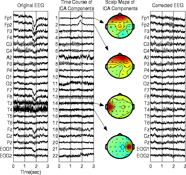
Now, let's see how this is applied in a real software package.
ICA applied to EEG part 8: Removing Artifactual Components in EEGLAB
The video 'ICA applied to EEG part 8: Removing Artifactual Components in EEGLAB' shows this exact process in the popular EEGLAB toolbox for MATLAB. This demonstrates the practical application of the theory.
Please watch the following segments: Identifying Eye and Muscle Components (1:03 - 2:58): Focus on how a component is identified as an artifact. Note the distinct features they look for: the scalp map (frontal for eyes), the frequency spectrum (1/f for eyes, high-power for muscle), and the ERPimage/time course (distinct spikes for blinks). Removing Components (4:46 - 7:04): Watch how the identified artifactual components are flagged for rejection and how the data is reconstructed. Pay attention to the 'before and after' plots that validate the cleaning process.
Other Advanced Methods
For completeness, you should be aware of other advanced methods, which are covered in the resources from Bitbrain and Mentalab (LINK, LINK). These include:
- Artifact Subspace Reconstruction (ASR): An automated method that finds clean periods of data and uses them to statistically remove noisy sections.
- Wavelet Decomposition: Breaks the signal down into constituent wavelets, allowing for artifactual components to be removed at specific time-frequency scales.
- Deep Learning: Newer methods use neural networks trained on large datasets to identify and remove artifacts automatically, a direction highly relevant to the cutting edge of neurotechnology.
Conclusion
Mastering artifact mitigation is an essential skill for anyone working seriously with EEG data. It transforms raw, noisy recordings into a clean signal that can be reliably analyzed and interpreted.
Key Takeaways:
- Artifacts are non-neural signals that can be physiological (EOG, EMG, ECG) or technical (60Hz, motion).
- Successful mitigation involves a three-stage strategy: proactive setup, careful recording procedures, and post-processing.
- Post-processing techniques range from simple filtering and epoch rejection to more sophisticated methods.
- Independent Component Analysis (ICA) is a powerful BSS technique that separates signals into independent sources, allowing for the targeted removal of artifacts like blinks and muscle activity while preserving valuable neural data.
- Identifying artifactual components in ICA involves examining their scalp topography, frequency spectrum, and time course.
Preview of the Next Lesson:
We have successfully designed our hardware and now know how to clean the data it produces. We finally have a signal we can trust! In our next lesson, "Apply spectral analysis (FFT) to identify EEG frequency bands (delta, theta, alpha, beta, gamma)," we will begin the exciting work of analyzing this clean signal to extract meaningful information about brain states. We'll use the Fast Fourier Transform (FFT) to break the EEG down into its fundamental frequency bands, which are the building blocks of cognitive and emotional state analysis.
Can't find a good explanation? Sign up and we'll make it for you
Sign up