
Hello again. Last time, you practiced the evidence needed to follow a neurite through a difficult region: trace from a confident anchor, compare alternative paths across several EM sections, and use the 3D reconstruction as a consistency check rather than proof.
Now you will convert that inspection into a useful diagnosis. When a reconstruction looks wrong, the practical question is not simply “is there an error?” It is: what kind of error is currently supported by the evidence? In this lesson, you will classify a candidate as a false split, false merge, missing branch, or uncertain case—without editing anything yet.
This is the classification vocabulary that will make later proofreading decisions safer and your eventual notes auditable.
Classification is about the reconstruction, not a single suspicious slice
An EM image shows biological material; a segmentation assigns that material to reconstructed objects. A candidate problem appears when those two disagree.
Keep three things separate:
- The biological question: Are these profiles physically part of the same neurite, different neurites, or a real branch point?
- The reconstruction question: Does the current selected reconstruction include the right material, omit correct material, or include foreign material?
- The evidence question: Can you demonstrate the conclusion across nearby sections, with membrane boundaries and local 3D morphology?
A useful engineering analogy is a system boundary bug:
- A false merge is like two independent objects accidentally sharing one identifier.
- A false split is like one object being broken into disconnected records.
- A missing branch is like discovering that an expected component is absent from the assembled result, without yet knowing exactly which record should be attached.
- Uncertain means the available observations do not justify a safe classification.
The labels describe what you currently know. They do not require you to predict the final edit or force a conclusion where the imagery is ambiguous.
The four classifications
1. False split: one neurite, incorrectly divided
A false split occurs when a single biological neurite is represented as two or more disconnected reconstructed pieces.
You should classify a candidate as a false split when you can identify:
- a selected branch that terminates or becomes disconnected;
- a separate candidate segment nearby;
- multi-section evidence that the two are physically continuous parts of the same membrane-bounded process.
The key word is identified. You are not merely saying “this neuron looks incomplete.” You have found a specific disconnected continuation and can trace why it belongs to the selected neurite.
Typical signs include:
- the selected segment stops at a location where the EM shows the neurite continuing;
- the plausible continuation is present as a separate segmentation color or object;
- a mitochondrion or other local landmark helps establish continuity across the gap;
- the two pieces form a coherent branch when considered in 3D, while the 2D EM confirms the membrane path.
A false split can be tiny—a broken thin twig—or substantial, such as a detached soma tract or a major dendritic extension.
Do not call it a false split merely because a branch seems short. If you cannot locate and validate the continuation, classify the situation as a missing branch or uncertain instead.
2. False merge: different neurites, incorrectly joined
A false merge occurs when the selected reconstruction includes material belonging to another neurite or cell. In other words, the current reconstructed object crosses a real membrane boundary that should separate two biological processes.
The diagnostic question is:
Does the selected object contain a connection that the EM evidence says is not biologically continuous?
Common visual patterns are useful alerts, although none replaces section-by-section inspection:
- X-shaped/perpendicular merger: two branches cross at roughly right angles and the segmentation treats the crossing as one connected object.
- H-shaped/parallel merger: two nearby parallel branches are joined by one or more short bridges.
- Twig-to-twig merger: thin terminal processes from otherwise separate arbors touch or are mistakenly connected.
- Synaptic-region merger: blurry or complex boundaries near a synapse cause the segmentation to combine distinct cells.
For a confident false-merge classification, find the actual erroneous bridge. In consecutive EM sections, you want evidence of two separately membrane-bounded processes that the current segmentation has joined.
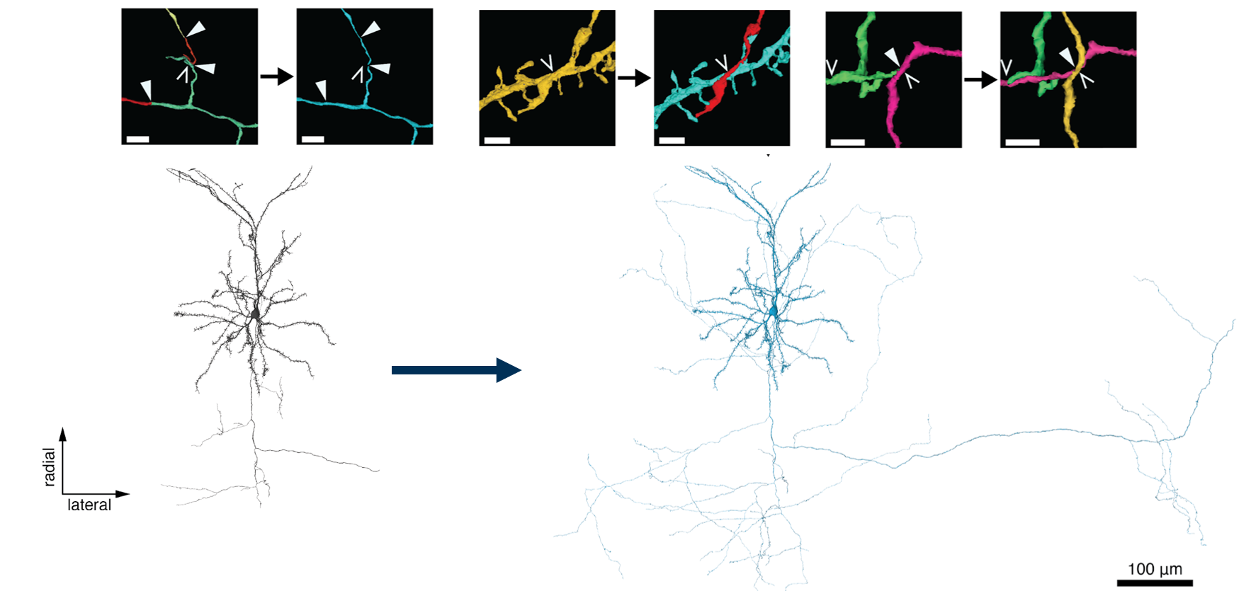
The figure highlights an important asymmetry:
- A false split removes correct material from a reconstruction by leaving it detached.
- A false merge adds incorrect material to a reconstruction by attaching a neighbor.
Both can make a neuron’s 3D form misleadingly plausible. That is why the EM stack remains decisive.
3. Missing branch: expected morphology, but no validated extension yet
A missing branch is a morphology-level observation: the displayed reconstruction appears to lack a branch that likely should be present.
Often, you first notice this in 3D. For example, an otherwise elaborate dendritic arbor has an unusually large empty wedge, or a broad parent branch appears to end abruptly without the expected continuation. The 3D view helps you notice the absence; it does not by itself prove that the absent branch exists.
Use missing branch when:
- the overall neuron morphology contains a suspicious gap, abrupt termination, or incomplete-looking region;
- you do not yet have enough local EM evidence to identify the exact disconnected continuation;
- the continuation may be absent from the current reconstruction, obscured by an artifact, or simply not yet found.
This category is deliberately different from a false split:
| Observation | Best classification |
|---|---|
| “This branch ends, but I can trace its separate continuation in EM.” | False split |
| “This arbor has a conspicuous void, but I have not located a specific continuation.” | Missing branch |
| “A branch might continue past an artifact, but several candidates remain plausible.” | Uncertain |
In practice, a missing branch may eventually be resolved as a false split once you locate the disconnected segment. Until then, “missing branch” accurately preserves what is known and what remains open.
4. Uncertain case: evidence is insufficient or contradictory
Uncertain is not a failure category. It is the correct classification when the image evidence cannot support a reliable decision.
Use it when:
- the target disappears into a fold, blackout, pinch, or severe misalignment;
- the membrane boundary cannot be resolved over enough consecutive sections;
- two or more continuations remain plausible after comparison;
- the 3D reconstruction suggests a problem but the 2D evidence does not locate it;
- a candidate depends mostly on expected cell morphology or on segmentation color, rather than direct image continuity.
A high-quality uncertain classification is specific about why it is unresolved. For example:
“Uncertain: selected twig ends before a folded section. Two nearby profiles emerge after the artifact, but neither can be connected through visible membrane continuity.”
That is far more valuable than guessing “false split” and creating an unsupported future edit target.
A focused reference: recurring merge and branch-loss patterns
The FlyWire Blog’s “FlyWire Proofreading Tips” gives a compact practical taxonomy of mergers, missing arbor regions, and false continuations. Read it now as a pattern-recognition reference, but keep the classification framework above in mind: visible pattern suggests where to inspect; multi-section EM evidence determines the label.
Read the relevant sections of “FlyWire Proofreading Tips” from the FlyWire Blog. It illustrates common false-merge patterns and explains why incomplete-looking morphology and path swaps require different kinds of inspection.
Start with “X-Shaped Mergers” and “H-Shaped or Parallel Mergers” in the opening section. Read the merger discussion, focusing on the difference between a crossing and a parallel bridge. Then read “Gaps in Dendritic Arbors” in the next section, from the holistic-arbor guidance. Notice that a 3D gap signals a candidate missing branch but does not itself identify the continuation. Finally, read “MIA Connections” and “Identifying Path Swaps” in the following section. Read the MIA-connection passage, then continue through the path-swap discussion. For this lesson, ignore the resource’s edit suggestions; focus on how ambiguous thin continuations and false continuations are recognized.
A conservative classification procedure
When you reach a suspicious site, apply the following procedure in order. This prevents the tempting but unreliable habit of labeling errors from a 3D silhouette alone.
Step 1: State what the selected reconstruction currently does
Describe only the observable reconstruction behavior:
- Does it stop abruptly?
- Does it include a branch that looks unrelated?
- Does it have a conspicuous void in its overall arbor?
- Does it become ambiguous because of image quality?
This first description should avoid conclusions. “Selected branch stops near ” is better than “split.”
Step 2: Inspect the local EM evidence
Return to a clear anchor before the suspicious region. Trace one section at a time and ask:
- Is there a membrane-bounded continuation of the selected neurite?
- Is it selected or disconnected?
- Are two membrane-bounded neurites being represented as one?
- Does the apparent connection remain visible across several sections?
- Is a global image shift or fold disrupting all nearby structures?
At this point, compare alternatives deliberately. If Candidate A and Candidate B are both plausible, trace both beyond the difficult slice before deciding.
Step 3: Use 3D to classify the morphology of the error
With the 2D hypothesis in mind, inspect the relevant portion of the reconstruction in 3D:
- A detached but plausible continuation supports a false split hypothesis.
- An extra branch creating an X, H, spur, or abrupt change in trajectory supports a false merge hypothesis.
- A conspicuous unfilled area in an otherwise coherent arbor supports a missing branch hypothesis.
- A smooth mesh that conceals unresolved 2D ambiguity should leave the case uncertain.
Step 4: Assign the most specific supported label
Use this priority order:
- False merge if foreign material is currently attached to the selected reconstruction and the erroneous connection is supported by EM evidence.
- False split if a specific disconnected continuation is identified as the same biological neurite.
- Missing branch if morphology strongly suggests absent material but no specific continuation is yet validated.
- Uncertain if the evidence remains inadequate, contradictory, or artifact-limited.
This is a reporting convention, not a claim that error mechanisms are always mutually exclusive.
The important mixed case: a path swap
A path swap occurs when a reconstruction begins on the correct neurite but then follows a nearby neurite instead. It is common where parallel processes travel closely together or where EM images are misaligned.
A path swap can contain two linked defects:
- the selected reconstruction has acquired the wrong neighboring continuation, which is a false merge;
- the true continuation is no longer part of the selected reconstruction, which may be a false split or a missing continuation.
For a single primary classification, call it false merge if the wrong material is currently attached and you can demonstrate the incorrect switch. Add a secondary qualifier in your scratch notes:
Primary: false merge (path swap).
Secondary: expected continuation is detached or not yet located.
This avoids an unsafe simplification. If you label the site only as a false split, a later reviewer might overlook the foreign branch that must also be addressed.
A useful diagnostic pattern is:
| Observation | Likely interpretation |
|---|---|
| Selected branch continues smoothly in 3D but changes physical trajectory in EM | Possible path swap |
| Two parallel neurites touch, then selected identity follows the neighbor | False merge, often path swap |
| Selected branch stops; nearby unselected process continues with membrane continuity | False split |
| Dendritic field has a large void but no candidate extension is confirmed | Missing branch |
| Folded or misaligned sections conceal the only possible connection | Uncertain |
Four short case walkthroughs
Case A: A branch stops beside a separate colored fragment
The selected branch approaches a dark mitochondrial oval and terminates. Across the next five sections, a nearby unselected segment surrounds the same membrane-bounded continuation and follows the original branch trajectory.
Classification: false split.
Why: there is a specific disconnected component, and multi-slice evidence supports one biological neurite.
Case B: An apparently X-shaped branch in 3D
In 3D, the selected reconstruction has a cross-like structure. In EM, a horizontal neurite and a vertical neurite remain separately membrane-bounded through the crossing region, but the segmentation includes both in the selected object.
Classification: false merge.
Why: two different neurites are currently represented as one reconstruction.
Case C: An asymmetric dendritic arbor
The 3D neuron has many branches around most of its body, but one large region contains a conspicuous gap. Several small nearby segments could be candidates, but none can yet be traced convincingly back to the selected arbor.
Classification: missing branch.
Why: the morphology indicates likely absent material, but a specific false split has not been established.
Case D: A thin branch reaches a damaged section
A thin selected neurite approaches a blackened or folded image region. After it, two similarly shaped processes appear. Neither can be followed with visible membrane continuity through the damaged area, and both fit the broad direction of travel.
Classification: uncertain.
Why: a “best guess” would not be evidence-based. Record the artifact and candidates rather than forcing a split classification.
A compact mental checklist
Before assigning a label, pause and ask:
- Foreign material attached? Check for a false merge.
- Specific detached continuation located? Check for a false split.
- Morphology suggests an absent branch, but continuation not located? Use missing branch.
- Image evidence blocked or alternatives unresolved? Use uncertain.
- Could this be a path swap? If yes, identify whether the selected reconstruction contains a wrong branch and note any missing true continuation separately.
For now, do not edit after classifying. Your goal is to make the diagnostic boundary clear: what is observed, what label the observation supports, and what remains unresolved.
Takeaways and next step
You can now classify a suspicious reconstruction site without conflating four distinct situations:
- A false split is a known disconnection within one biological neurite.
- A false merge is an incorrect attachment of different neurites in one reconstruction.
- A missing branch is a morphology-based indication of likely absent material when the exact continuation is not yet validated.
- An uncertain case is the correct result when artifacts or ambiguous EM evidence prevent a defensible conclusion.
Remember that path swaps often combine a false merge with a missing or detached correct continuation. Classify the foreign attached material as the primary local problem, then record the related missing path separately.
Next, you will turn these classifications into concise candidate-problem notes containing location, visual evidence, uncertainty, and a proposed next step.
Can't find a good explanation? Sign up and we'll make it for you
Sign up