
Hello! Welcome to the final lesson in our module on osseointegration.
In our previous lesson, we established the critical difference between successful osseointegration and failure via fibrous encapsulation. We saw that failure isn't a passive process, but an active biological response—scar formation—often triggered by factors like micromotion that disrupt the delicate healing environment at the implant interface.
Today, we will broaden this analysis. Your learning outcome is to analyze how systemic and local factors (e.g., smoking, diabetes, infection, micromotion) can disrupt the biological cascade and inhibit successful osseointegration. We will systematically investigate the key culprits that can derail the journey from a placed implant to a stable, integrated foundation, connecting these clinical risk factors back to the cellular and molecular machinery we've studied.
A Framework for Failure: Local vs. Systemic Factors
The success of osseointegration hinges on a multitude of variables. A useful way to organize them is by their scope of influence:
- Local Factors: Conditions specific to the implant site itself. These include the quality and quantity of the host bone, the surgical technique, and the immediate mechanical and biological environment of the implant.
- Systemic Factors: Conditions that affect the patient's entire body and, consequently, their overall capacity for healing. These encompass metabolic diseases, medications, and lifestyle choices.
Essentially, for osseointegration to succeed, we need both a suitable local environment and a host system that is primed for effective regeneration. Let's dissect how disruptions in each of these domains can lead to failure.
Local Factors: The Immediate Battleground
These factors act directly at the bone-implant interface, influencing the initial cellular events.
1. Micromotion: The Mechanical Saboteur
We concluded our last lesson by identifying micromotion as a primary trigger for fibrous encapsulation. Now, let's quantify this relationship. Research shows a clear threshold: if movement between the implant and bone is too high during the initial healing phase, the delicate process of bone formation is physically disrupted.
- The initial fibrin clot, which acts as a scaffold for migrating cells, is torn apart.
- The formation of new, fragile blood vessels (angiogenesis) is inhibited, starving the site of oxygen and nutrients.
- The mechanical strain on mesenchymal stem cells steers their differentiation toward fibroblasts (scar-forming cells) rather than osteoblasts (bone-forming cells).
The images below, from a systematic review on the topic, powerfully illustrate this dose-response relationship.
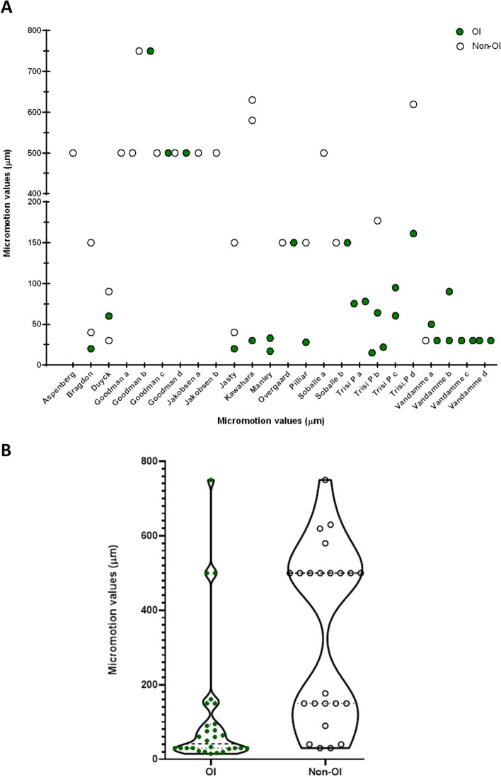
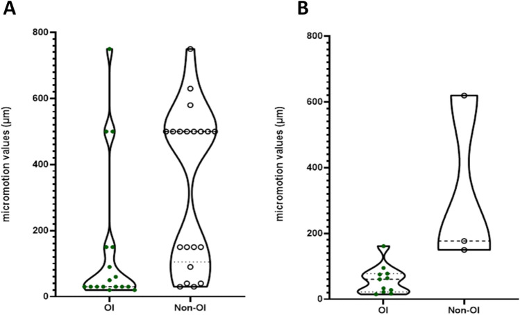
From your background in data analysis, you can see that a violin plot shows the probability density of the data. The narrow "waist" and tight distribution for the osseointegration group indicate a strict requirement for mechanical quietude. In clinical terms, this underscores the critical importance of achieving high primary stability at the time of surgery.
2. Infection: The Biological Saboteur
While the implant surgery is performed under sterile conditions, the oral cavity is home to a complex microbiome. If bacteria contaminate the implant site during or shortly after surgery, they can initiate a cascade that is directly hostile to bone formation.
To see what this looks like clinically and understand its causes, let's watch a short video.
Failing Dental Implant? What causes it? How do I Treat It? Does it Need to be Removed?
Dr. David Choi, in the video 'Failing Dental Implant?', discusses the common causes of implant failure, focusing on the role of infection (peri-implantitis) and mechanical trauma.
Please watch from 02:38 to 04:20. Pay attention to his explanation of how bacterial infection, cement leakage, and excessive trauma contribute to bone loss around an implant.
As Dr. Choi explains, bacterial contamination triggers an aggressive inflammatory response. This connects directly to the concept of immunomodulation we've touched on previously.
- The Immune Shift: A healthy healing response involves a transition from a short, pro-inflammatory phase (dominated by M1 macrophages) to a longer, pro-regenerative phase (dominated by M2 macrophages). A persistent bacterial infection traps the immune system in the pro-inflammatory M1 state. These cells release cytokines that, instead of promoting bone formation, actively recruit osteoclasts and promote bone resorption.
- Hostile Environment: The bacterial biofilm can create a localized, acidic microenvironment that is detrimental to osteoblast function and survival.
This process, where infection leads to inflammation and subsequent bone loss around an implant, is termed peri-implantitis. While it's a major cause of long-term failure, an acute infection during the initial healing phase will prevent osseointegration from ever occurring, leading directly to fibrous encapsulation.
Systemic Factors: Compromising the Host
Systemic factors don't necessarily target the implant site itself, but they degrade the body's overall ability to perform the complex task of bone regeneration.
Let's begin with a clinician's perspective on the major systemic red flags.
The Top Reasons You Should NOT Get Dental Implants
In the video 'The Top Reasons You Should NOT Get Dental Implants', Dr. Brett Langston provides a practical overview of several key risk factors.
Watch the segments on uncontrolled diabetes (01:08 - 01:38) and smoking (04:08 - 05:08). These provide the clinical context for our deeper biological dive.
Now, let's unpack the mechanisms behind these clinical observations, using a high-level review paper that is well-suited to your preference for detailed sources.
Osseointegration—the biological reality of successful dental ...
The paper 'Osseointegration—the biological reality...' by Cooper and Shirazi provides an excellent, in-depth discussion of how systemic conditions can disturb the healing process at a molecular level.
Please read the sections 'Osseointegration re-defined' and 'Osseointegration disturbed'. The first section discusses the critical role of the immune system (immunomodulation) in successful healing. The second directly addresses how systemic diseases and medications interfere with this process.
Drawing from the video and the paper, let's analyze the mechanisms for two major systemic factors.
1. Uncontrolled Diabetes Mellitus
As Dr. Langston noted, this is a major concern. The problem is multifaceted, stemming from the systemic effects of hyperglycemia:
- Impaired Circulation (Microangiopathy): High blood sugar damages small blood vessels, reducing blood flow to the surgical site. This impairs the delivery of oxygen, nutrients, growth factors, and immune cells necessary for healing.
- Dysfunctional Immune Response: Hyperglycemia impairs the function of neutrophils and macrophages, reducing their ability to fight off infection and to transition from a pro-inflammatory to a pro-regenerative state. This leaves the patient more susceptible to infection and stuck in a state of chronic inflammation.
- Advanced Glycation End-products (AGEs): These molecules form when excess sugar binds to proteins like collagen. The accumulation of AGEs in bone tissue makes the matrix stiffer, more brittle, and directly suppresses the function of osteoblasts, while promoting osteoclast activity.
In essence, diabetes creates a system-wide environment that is simultaneously prone to infection, deficient in circulation, and biochemically biased against bone formation.
2. Smoking
Smoking introduces a cocktail of chemicals that systematically undermines healing:
- Vasoconstriction & Hypoxia: Nicotine is a potent vasoconstrictor, acutely reducing blood flow to peripheral tissues, including the jawbone. Furthermore, carbon monoxide from smoke binds to hemoglobin with much higher affinity than oxygen, reducing the blood's oxygen-carrying capacity. The result is a hypoxic (oxygen-starved) healing environment.
- Direct Cellular Toxicity: Nicotine and other toxins in tobacco smoke have been shown to directly inhibit the proliferation and function of osteoblasts.
- Impaired Immune Function: Similar to diabetes, smoking impairs the function of the immune cells required for a clean and effective healing process.
- Physical Disruption: As Dr. Langston mentioned, the negative pressure created by the act of smoking can physically dislodge the initial blood clot, which is the foundational scaffold for healing.
3. Other Factors
The Cooper & Shirazi paper also highlights other systemic disruptors:
- Medications: Drugs like bisphosphonates (for osteoporosis), while designed to prevent bone loss, do so by shutting down bone turnover. This can interfere with the dynamic remodeling required for osseointegration. Other common medications like SSRIs (antidepressants) and proton pump inhibitors (for acid reflux) have also been statistically linked to higher implant failure rates, likely through off-target effects on bone metabolism.
- Irradiated Bone: Patients who have undergone radiation therapy for head and neck cancers have compromised bone with reduced vascularity and cellularity, making osseointegration extremely challenging.
Test your understanding!
A patient has two failing implants placed six months ago. Implant A, in an area of soft bone, is mobile and an X-ray reveals fibrous encapsulation. Implant B, in a patient who is a heavy smoker, never fully healed and developed a persistent, low-grade infection.
Which fundamental principle of osseointegration was most likely violated for Implant A? And for Implant B, which two disruptive factors are likely acting synergistically?
Show answer
For Implant A, the most likely violated principle is primary stability. Soft bone provides poor initial mechanical grip, leading to micromotion that exceeded the biological tolerance, resulting in fibrous encapsulation.
For Implant B, the two factors acting synergistically are a local factor (infection) and a systemic factor (smoking). Smoking created a systemically compromised healing environment (poor circulation, impaired immunity), making the site much more vulnerable to the local bacterial challenge, which in turn drove a persistent inflammatory response that prevented bone formation.
Conclusion
In this final lesson of the module, we have analyzed the key factors that can disrupt osseointegration, categorizing them into local and systemic influences.
Key Takeaways:
- Successful osseointegration requires both a favorable local environment and a competent systemic host response.
- Local factors like micromotion (due to poor primary stability) and infection directly subvert the biological cascade at the implant site, promoting scar formation and bone resorption.
- Systemic factors like uncontrolled diabetes and smoking degrade the body's overall healing capacity by impairing circulation, compromising the immune response, and directly inhibiting the cells responsible for bone formation.
- Ultimately, all these factors disrupt the delicate balance of the healing cascade, tipping it away from regeneration (osteogenesis) and toward a state of chronic inflammation and/or fibrosis.
Next Lesson Preview:
Understanding these risk factors is the foundation of modern implant dentistry. It's not enough to know what can go wrong; the goal is to prevent it from happening. In our next module, "Pre-Surgical Assessment and Digital Planning," we will transition from biology to clinical practice. We'll begin by exploring how clinicians use advanced imaging tools, specifically Cone Beam Computed Tomography (CBCT), to meticulously evaluate the patient's anatomy and bone quality, which is the first and most critical step in mitigating many of the local risk factors we have discussed today.
Can't find a good explanation? Sign up and we'll make it for you
Sign up