
Hello! Welcome to your lesson on Electrooculography.
In our last session, we delved into Heart Rate Variability (HRV) and how it reveals the dynamic balance of the autonomic nervous system. We saw how metrics like RMSSD and High-Frequency power provide a powerful window into parasympathetic activity and overall physiological stress.
Today, we'll explore another crucial biosignal that's highly relevant to your work at Neuraease. This lesson addresses the learning outcome: Describe the principles of Electrooculography (EOG) for tracking eye movements and detecting blinks. While EOG has many applications, its most critical role in a neurotechnology context is for identifying and removing eye-movement artifacts from EEG data—a fundamental step for reliable analysis.
1. The Physiological Principle of EOG
The ability to measure eye movements with simple electrodes stems from a fascinating property of the eye itself. The front of the eye (the cornea) is electrically positive relative to the back of the eye (the retina). This creates a constant electrical potential difference, known as the corneo-retinal standing potential, which is typically in the range of 0.4 to 1.0 mV.
This potential turns the eye into a small biological dipole, with the positive pole pointing forward along the optical axis.
Electrooculogram - Recording Eye Movements with the Heart and Brain SpikerShield
The video from Backyard Brains provides a very clear and concise explanation of this dipole concept.
Watch the short clip from 01:10 to 01:29. It perfectly visualizes how the eye acts as a dipole and how its movement changes the surrounding electric field.
When you place electrodes on the skin around the eyes, they can detect the changes in this electric field as the eye rotates.
- When the cornea (positive pole) moves toward an electrode, the potential at that electrode increases.
- When the cornea moves away from an electrode, the potential decreases.
This principle is the foundation of EOG.
Electrooculograms for Human–Computer Interaction
For a more detailed description of this process, let's turn to a scientific review paper on EOG. It clearly explains the origin of the signal.
Please read the first paragraph of Section 2, 'Characteristics'. It begins with 'An EOG is a signal that changes according to eye-movements...' and ends just before Figure 1. Pay attention to how the potential changes as the cornea approaches or moves away from an electrode.
2. How to Measure EOG Signals
To capture these potential changes, we use a differential measurement setup with at least two channels: one for horizontal movements and one for vertical movements.
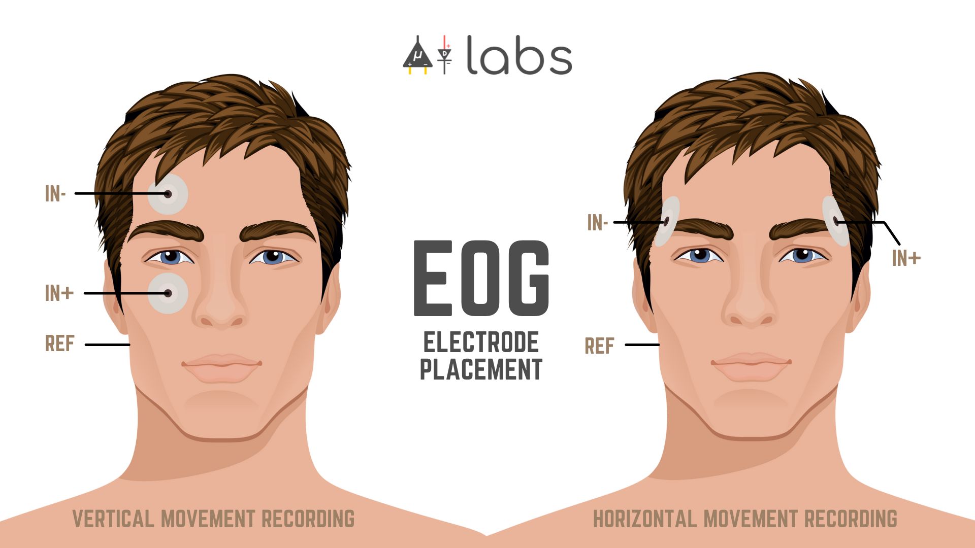
- Horizontal Channel: Electrodes are placed at the outer corners (the outer canthus) of the left and right eyes. The potential difference between these two electrodes will primarily reflect left-right eye movements.
- Vertical Channel: Electrodes are placed above and below one eye (typically the same eye used for the horizontal channel). The potential difference here will reflect up-down eye movements and, very importantly, eye blinks.
Electrooculogram - Recording Eye Movements with the Heart and Brain SpikerShield
Let's see this in practice. The Backyard Brains video demonstrates the electrode setup for both channels.
First, watch from the beginning until 01:10 to see the setup for recording horizontal (left vs. right) movements. Then, watch from 02:16 to 02:45 to see the setup for the vertical (up vs. down) channel.
3. Interpreting EOG Waveforms
The differential signals from the horizontal and vertical channels produce distinct waveforms corresponding to different eye movements and blinks.
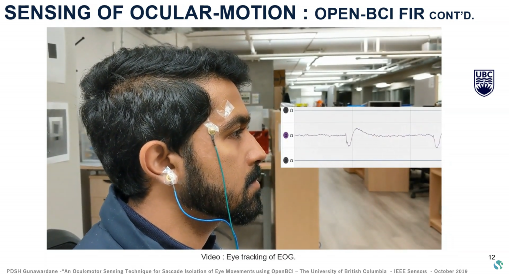
Eye Movement Signatures
The polarity of the signal deflection depends on which electrode is connected to the positive or negative input of your amplifier, but the pattern is consistent. Assuming a standard configuration:
- Looking Right: The cornea moves toward the right electrode, creating a positive deflection in the horizontal channel.
- Looking Left: The cornea moves away from the right electrode, creating a negative deflection.
- Looking Up: The cornea moves toward the upper electrode, creating a positive deflection in the vertical channel.
- Looking Down: The cornea moves toward the lower electrode, creating a negative deflection in the vertical channel.
Electrooculography ( EOG) - Basics of Biomedical
This video clearly illustrates the expected waveforms for each primary direction of eye movement.
Watch the segment from 08:13 to 09:44. It shows the distinct EOG waveforms generated by left, right, up, and down eye movements, along with typical voltage ranges.
The Signature of an Eye Blink
Eye blinks produce a very distinct, sharp, high-amplitude pulse that is most prominent in the vertical channel. This happens because as the eyelid moves down over the cornea, it acts like a sliding conductor, momentarily connecting the positive cornea to the skin surface and causing a large, rapid change in potential.
A typical spontaneous blink has:
- Amplitude: 100 - 400 µV (often much larger than signals from eye rotation).
- Duration: 200 - 400 ms.
- Shape: A rapid upward spike (lid closing) followed by a slightly slower downward recovery (lid opening).
EOG Signals in Drowsiness Research
The paper on 'EOG Signals in Drowsiness Research' provides an excellent description of the blink signal.
Please read the section '2.4.2 Blink Signal and Variables' and the short section '4.3 Normal blink'. These sections describe the characteristic amplitude, duration, and shape of a blink as seen in an EOG signal.
Test your understanding!
Imagine you are looking at two EOG channels from a user. You observe the following sequence of events:
- A large, sharp, positive pulse appears mainly in the vertical channel.
- A slow, rectangular positive pulse appears in the horizontal channel, lasting for about 1 second.
- A slow, rectangular negative pulse appears in the vertical channel, lasting for about 1 second.
What did the user most likely do?
Show answer
- Event 1 (Sharp vertical pulse): The user blinked.
- Event 2 (Slow horizontal pulse): The user looked to the right and held their gaze for one second.
- Event 3 (Slow vertical pulse): The user looked down and held their gaze for one second.
4. Application: Blink Detection and Artifacts
Understanding the EOG signal is crucial for two main reasons in your field:
- As a Signal: Eye movements and blinks can be used as features to infer cognitive states like drowsiness, attention, or as commands for a Human-Computer Interface (HCI).
- As Noise: Eye movements and blinks are one of the largest sources of contamination (artifacts) in EEG recordings. If not removed, they can completely obscure the underlying brain activity.
A simple way to detect blinks is to apply a threshold to the vertical EOG channel. Since blinks have a much higher amplitude than other eye movements, any signal that crosses a predefined voltage level can be classified as a blink.
Electrooculogram - Recording Eye Movements with the Heart and Brain SpikerShield
The Backyard Brains video concludes with a great demonstration of a simple threshold-based blink detector.
Watch from 03:01 to 03:35. This shows how easily a blink can be detected in real-time using a simple voltage threshold, demonstrating a practical application of the principles we've discussed.
While simple thresholding works, more advanced algorithms are used in practice for higher accuracy. It's also vital to be aware of other movements that can create artifacts in the EOG signal, such as yawning, talking, and head movements. Being able to distinguish these from true eye movements and blinks is a key challenge in real-world data analysis.
Conclusion
You have now learned the fundamental principles of Electrooculography. You can describe how it works, how it's measured, and how to interpret the resulting signals for basic eye movements and blinks.
Key Takeaways:
- Principle: EOG works by measuring the corneo-retinal potential, which makes the eye an electrical dipole.
- Measurement: A horizontal channel tracks left-right movements, while a vertical channel tracks up-down movements and blinks.
- Signatures: Eye rotations produce slower, rectangular deflections, while blinks produce sharp, high-amplitude spikes in the vertical channel.
- Application: EOG is used to track eye movements and detect blinks, which can be both a valuable signal for assessing user state and a major source of artifacts in EEG recordings.
Preview of the Next Lesson:
In our next and final lesson of this module, we will tie everything together. You will be asked to propose a multimodal sensor configuration to assess stress or emotional state. Building on your knowledge of EDA, HRV, and now EOG, you will think like a system designer and decide which sensors to combine to create a robust system for your startup, Neuraease.
Can't find a good explanation? Sign up and we'll make it for you
Sign up