
Hello! Welcome to the second module of your course on dental implantation.
In the first module, we established a solid understanding of the macroscopic anatomy of the jawbones and surrounding structures. Now, we will zoom in from the organ level to the cellular level. The success of a dental implant is fundamentally a biological process, and to understand it, we must first understand how bone lives, adapts, and heals.
This lesson addresses the first learning outcome of Module 2: Explain the specific roles of osteoblasts, osteoclasts, and osteocytes in the continuous process of bone metabolism. We will explore the three key cell types that govern the life of bone tissue, treating them as a dynamic, interconnected system responsible for maintaining the skeleton's integrity.
Understanding these cellular actors is the bedrock for grasping concepts like osseointegration, bone grafting, and the factors that determine implant success, which we will cover in subsequent lessons.
1. The Dynamic Nature of Bone
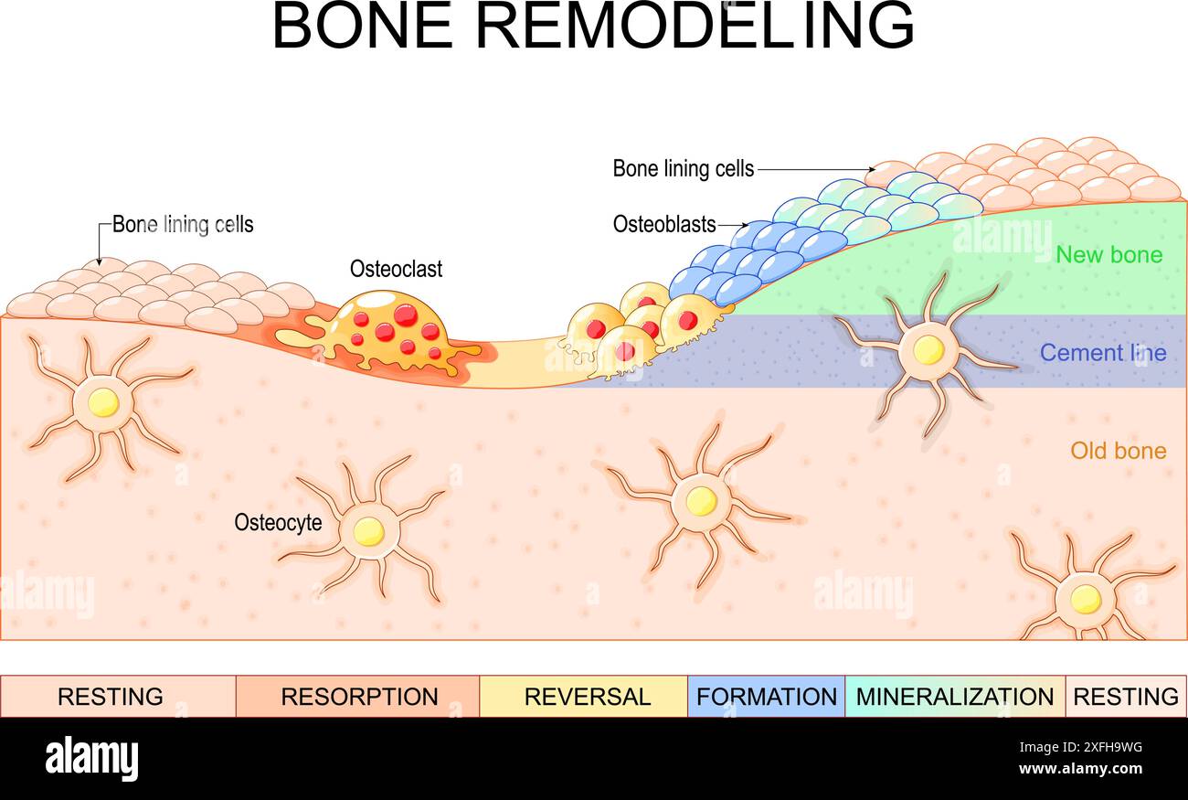
Contrary to its appearance as a static, inert material, bone is a highly dynamic and metabolically active tissue. It is in a constant state of renewal through a process called bone remodeling. This involves the coordinated removal of old or damaged bone and its replacement with new, healthy bone. This process is essential for repairing micro-damage from daily activities, adapting to mechanical loads, and maintaining calcium homeostasis in the body.
The key players in this process are three specialized cells. Let's get a quick overview of the team.

To see how these cells work together, please watch the following video. It provides an excellent visual introduction to the remodeling cycle.
This video from Osmosis from Elsevier provides a clear, animated overview of the bone remodeling process, introducing the main cells and their basic interactions.
Please watch the following segments to get a conceptual framework for our lesson: Introduction to Remodeling (0:04 - 0:31) Osteoblast and Osteoclast Interaction (2:50 - 4:06) Bone Formation (4:20 - 4:49) Summary (5:50 - 6:25) Focus on the sequence of events: how resorption is initiated and how formation follows.
As the video illustrates, bone remodeling is a beautifully orchestrated dance between destruction and creation. Now, let's delve into the specific roles and characteristics of each of these cellular architects.
2. The Cellular Players in Detail
Your preference for in-depth, primary sources will be well-served by the following review article. We will use it to build a detailed picture of each cell type.
A Brief Review of Bone Cell Function and Importance - PMC
We will now refer to the paper 'A Brief Review of Bone Cell Function and Importance' from the journal Cells. It provides a comprehensive, peer-reviewed look at the physiology of bone cells.
For now, just be aware of this resource. We will read specific sections as we discuss each cell type below.
2.1. Osteoclasts: The Resorption Specialists
Osteoclasts are the cells responsible for breaking down bone tissue, a process called resorption. They are large, multinucleated cells that originate not from the local bone cell lineage, but from hematopoietic stem cells in the bone marrow—the same lineage that produces monocytes and macrophages.
Please read the section on osteoclasts in the review paper.
A Brief Review of Bone Cell Function and Importance - PMC
This section of the review paper details the origin, morphology, and function of osteoclasts.
Please read the subsection titled 'Osteoclasts' within Section 2.2.3, 'Cells of Bone'. You can find this by searching for the heading 'Osteoclasts'. Focus on understanding their origin, their multinucleated nature, and the function of the 'ruffled border' and 'sealing zone'.
To perform resorption, an osteoclast attaches firmly to the bone surface, creating a sealed microenvironment underneath it. This is achieved by the sealing zone, an adhesive ring of proteins. Within this sealed compartment, the cell membrane is highly folded into a ruffled border, which dramatically increases its surface area.
The osteoclast then acts as a powerful biological machine:
- Acidification: It pumps protons (H⁺ ions) across the ruffled border, creating a highly acidic environment (pH ≈ 4.5). This acid dissolves the inorganic mineral component of the bone, hydroxyapatite ().
- Enzymatic Digestion: It secretes enzymes, most notably Cathepsin K, into the compartment. These enzymes digest the organic matrix, which is primarily composed of collagen.
The degraded components are then endocytosed by the osteoclast, transported across the cell, and released into the bloodstream. Once its task is complete, the osteoclast undergoes apoptosis (programmed cell death).
2.2. Osteoblasts: The Master Builders
In direct opposition to osteoclasts, osteoblasts are the cells responsible for synthesizing new bone tissue. They originate from mesenchymal stem cells, which are part of the connective tissue lineage.
Now, please read the corresponding section on osteoblasts in the review paper.
A Brief Review of Bone Cell Function and Importance - PMC
This section describes osteoblasts, the bone-forming cells.
Please read the subsection titled 'Osteoblasts' within Section 2.2.3. Focus on their origin, their primary function of secreting osteoid, and the two-step process of bone formation (matrix deposition and mineralization).
The function of osteoblasts is a two-stage process, analogous to creating reinforced concrete:
- Osteoid Deposition: Osteoblasts first secrete the unmineralized organic matrix of bone, called osteoid. This is about 90% Type I collagen, which forms a protein scaffold that gives bone its tensile strength and flexibility (the "rebar").
- Mineralization: Following osteoid deposition, osteoblasts orchestrate the mineralization process. They release vesicles containing calcium and phosphate, which nucleate to form hydroxyapatite crystals. These crystals grow and deposit within the collagen scaffold, giving bone its hardness and compressive strength (the "concrete").
Once they have finished forming bone in a particular location, mature osteoblasts have one of three fates:
- They become entrapped in the matrix they just created and differentiate into osteocytes.
- They flatten and become quiescent bone lining cells that cover the bone surface.
- They undergo apoptosis.
2.3. Osteocytes: The Command and Control Network
Osteocytes are the most abundant cell type in mature bone, making up 90-95% of all bone cells. They are terminally differentiated cells that were once osteoblasts.
Please read the final section on osteocytes.
A Brief Review of Bone Cell Function and Importance - PMC
This section covers osteocytes, the most numerous and long-lived bone cells.
Please read the subsection titled 'Osteocytes'. Pay close attention to how they are formed, their dendritic morphology, and their proposed role as mechanosensors within the lacuno-canalicular network.
Osteocytes reside in small spaces within the mineralized matrix called lacunae. They are not isolated; they extend long, dendritic processes through tiny channels called canaliculi to form a vast, interconnected network. This network allows them to communicate with each other, with the cells on the bone surface (osteoblasts and lining cells), and with the blood supply.
The primary role of the osteocyte is that of a mechanosensor and orchestrator. Given your background in physics and modeling, you can think of the osteocyte network as a distributed, living sensor array embedded within a material.
- Sensing: When bone is subjected to mechanical load, the fluid within the canaliculi flows, creating shear stress on the osteocyte processes. The cells sense this physical stimulus.
- Orchestration: In response to these mechanical signals, osteocytes release signaling molecules that direct the activity of osteoblasts and osteoclasts on the bone surface. They can call for bone to be added in areas of high strain or removed from areas of low strain. This is the cellular basis of Wolff's Law, which we will discuss in the next lesson.
3. The Regulatory Axis: How Bone Cells Communicate
The balance between bone resorption and formation is not random; it is tightly regulated. The primary signaling pathway that governs this balance is the RANK/RANKL/OPG system. This system is a perfect example of how osteoblasts and osteocytes control the activity of osteoclasts.
To understand this crucial regulatory axis, please watch the following focused video.
Introduction to RANKL | RANK | OPG Signaling Pathway
This video from JJ Medicine provides a more detailed explanation of the RANK/RANKL/OPG signaling pathway, which is central to the regulation of bone resorption.
Please watch the first two sections of the video: Introduction to RANKL, RANK, OPG (0:00 - 2:24) Regulation of Osteoclast Differentiation and Activation (2:07 - 4:35) Focus on identifying which cells produce each of the three key proteins (RANK, RANKL, OPG) and what the outcome of their interaction is.
Here is a summary of this critical control system:
- RANKL (Receptor Activator of Nuclear factor κβ Ligand): This protein is the primary "go" signal for osteoclast formation and activation. It is produced by osteoblasts and osteocytes.
- RANK (Receptor): This is the receptor for RANKL. It is expressed on the surface of osteoclast precursors and mature osteoclasts. When RANKL binds to RANK, it triggers the differentiation of precursors into mature osteoclasts and activates them to begin resorption.
- OPG (Osteoprotegerin): This protein is the "stop" signal. It is also produced by osteoblasts and osteocytes. OPG acts as a decoy receptor, binding to RANKL and preventing it from binding to RANK. This inhibits osteoclast formation and activation.
The ratio of RANKL to OPG produced by osteoblasts and osteocytes essentially functions as a rheostat for bone resorption.
- High RANKL/OPG ratio → More RANKL available to bind RANK → Increased osteoclast activity → Net bone resorption.
- Low RANKL/OPG ratio → Most RANKL is neutralized by OPG → Decreased osteoclast activity → Net bone formation (or stasis).
This elegant system ensures that the cells building the bone (osteoblasts) and sensing its needs (osteocytes) are the ones that ultimately control the cells that demolish it (osteoclasts).
Conclusion
In this lesson, we have dissected the cellular machinery that drives the continuous process of bone metabolism.
Key Takeaways:
- Bone is a dynamic tissue constantly being remodeled by a team of specialized cells.
- Osteoclasts, derived from hematopoietic stem cells, are responsible for bone resorption. They create a sealed, acidic compartment to dissolve mineral and digest the organic matrix.
- Osteoblasts, derived from mesenchymal stem cells, are responsible for bone formation. They build a collagen-based scaffold (osteoid) and then mineralize it.
- Osteocytes are former osteoblasts that become embedded in the bone matrix. They form a vast sensor network that detects mechanical strain and orchestrates the remodeling process by directing the activity of osteoblasts and osteoclasts.
- The communication between these cells is critical. The RANK/RANKL/OPG axis is a key regulatory pathway where osteoblasts and osteocytes control osteoclast activity, thereby coupling bone formation and resorption.
Preview of the Next Lesson:
We have just scratched the surface of bone regulation by focusing on the RANK/RANKL/OPG pathway. In our next lesson, we will broaden our view to describe the full biochemical signaling cascade involved in bone remodeling and the many other factors that regulate it, including systemic hormones (like parathyroid hormone and estrogen) and local growth factors. This will provide a more complete picture of how the body maintains skeletal health.
Can't find a good explanation? Sign up and we'll make it for you
Sign up