
Hello! Welcome to the next lesson in our course on dental implantation.
In our last session, we detailed the complex biochemical signaling network that governs bone remodeling, including the key roles of the RANKL/OPG axis, Wnt signaling, and various growth factors. We now have a map of the cellular machinery. Today, we will investigate the primary physical stimulus that directs this machinery.
This lesson addresses the learning outcome: Explain Wolff's law and how mechanical loading and strain distribution influence bone density and architecture.
We will begin by defining Wolff's Law and exploring its historical origins. Then, we will delve into the modern understanding of mechanotransduction—the process by which bone cells, particularly osteocytes, sense physical forces and convert them into the biochemical signals we discussed previously. Finally, we'll examine the consequences of this process, including how it shapes bone architecture and the critical clinical concept of stress shielding.
Given your background in physics and numerical simulation, this lesson's focus on the interplay between mechanical forces, strain, and biological response should be particularly relevant.
1. Wolff's Law: Form Follows Function
At its core, Wolff's Law is a simple and elegant principle that describes the adaptive nature of bone. Let's start with a clear definition and some intuitive examples.
This short video from Explified Labs provides a concise introduction to Wolff's Law and illustrates it with several accessible examples.
Please watch the first two sections of the video, 'Introduction to Wolff's Law' and 'Examples of Wolff's Law in Action' (0:00 - 1:48). Focus on the core statement of the law and how it manifests in different scenarios, like in athletes and astronauts.
As the video explains, Wolff's Law, articulated by the German surgeon Julius Wolff in the 19th century, states that bone remodels itself to adapt to the mechanical stresses it experiences. Increased load leads to stronger, denser bone, while decreased load leads to weaker, less dense bone.
This idea didn't arise in a vacuum. It was the culmination of centuries of anatomical observation. To appreciate this, let's briefly look at the historical context.
Evolving concepts in the mechanobiology of bone
The article 'Evolving concepts in the mechanobiology of bone' by Murray C. Meikle provides an excellent historical account. We will look at the section that builds up to Wolff's work.
Please read the sections titled 'The trajectoral theory of bone' and 'Julius Wolff and his law'. This will give you insight into the collaboration between anatomist von Meyer and engineer Culmann, which laid the groundwork for Wolff's famous law.
The "trajectoral theory" is a beautiful example of cross-disciplinary insight, combining anatomy and engineering. The observation that the internal trabeculae of the femur align with the stress trajectories of a similarly shaped crane provided a powerful mechanical explanation for bone's internal architecture.

Wolff synthesized these observations into his "Law of Bone Transformation." However, as the reading notes, Wolff's own understanding of the underlying physiology was limited. The how of this adaptation remained a black box for nearly a century.
2. Mechanotransduction: How Bone Senses Load
Wolff's Law describes a phenomenon at the organ level. The modern challenge is to explain it at the cellular and molecular level. The process by which cells convert mechanical stimuli into electrochemical activity is called mechanotransduction. In bone, the primary mechanosensors are the osteocytes embedded within the mineralized matrix.
The following review paper provides a detailed, graduate-level exploration of this topic.
The paper 'Boning up on Wolff's Law' from the Journal of Biomechanics is a comprehensive resource that details our modern understanding of bone mechanobiology. We will use it to break down the process of mechanotransduction.
First, please read Section 2, 'Forces experienced by bone cells'. This section is particularly relevant to your physics background, as it describes how macroscopic loads are translated into microscopic forces. Next, read the introduction to Section 4 and Section 4.1, 'Osteocyte mechanosensing and mechanotransduction'. This explains the cellular machinery that detects these forces.
Let's synthesize the key points from that reading.
2.1. The Physical Stimuli at the Cellular Level
A macroscopic load on a bone (e.g., from walking) doesn't just strain the bulk material. It creates a complex mechanical environment for the osteocytes within their lacunae. The primary stimuli are:
- Interstitial Fluid Flow: As the bone matrix deforms, it squeezes the interstitial fluid through the network of canaliculi. This creates fluid shear stress on the osteocyte cell membranes and their processes. This is analogous to transport phenomena in a porous medium. The oscillatory nature of loads like walking creates corresponding oscillatory fluid flow, which appears to be a potent stimulus.
- Hydrostatic Pressure: The compression of the fluid within the lacunar-canalicular system generates pressure changes.
- Direct Matrix Strain: The mineralized matrix itself deforms, and this strain can be amplified at the local level around the lacunae, directly stretching the osteocytes and their connections to the matrix.
2.2. The Osteocyte's Sensory Toolkit
The osteocyte uses several mechanisms, described in the paper, to detect these physical forces:
- The Cytoskeleton and Integrins: Integrins are transmembrane proteins that anchor the cell's internal cytoskeleton to the extracellular matrix. Strain and fluid drag forces are transmitted through these connections, triggering intracellular signaling.
- Primary Cilia: These single, non-motile cilia act like antennae, bending in response to fluid flow and activating signaling pathways.
- Ion Channels: Mechanosensitive ion channels open in response to membrane stretch, altering the cell's membrane potential and allowing influx of ions like Ca²⁺, a key second messenger.
- Hemichannels/Gap Junctions: These channels allow communication between osteocytes and can release signaling molecules like ATP and PGE₂ into the extracellular space in response to mechanical stimulation.
2.3. Closing the Loop: From Force to Biochemistry
This is where we connect today's lesson to the previous one. Once an osteocyte senses a mechanical load, it adjusts its production of the very signaling molecules we've already discussed. As noted in Section 4.2 of the "Boning up on Wolff's Law" paper, mechanical loading typically leads to:
- Decreased RANKL/OPG Ratio: Osteocytes reduce their expression of RANKL, tipping the balance away from osteoclast activation.
- Decreased Sclerostin: Sclerostin is a powerful inhibitor of the anabolic Wnt pathway. By reducing sclerostin production, osteocytes "release the brakes" on osteoblasts, promoting bone formation.
In essence, mechanical loading tells the osteocytes to send out signals that say: "Inhibit resorption and promote formation."
3. Consequences: Bone Adaptation and Clinical Relevance
The net effect of this mechanotransduction system is the adaptation of bone mass and architecture to its functional demands.
3.1. Bone Gain and the Mechanostat Theory
Vigorous physical activity increases bone density. This is Wolff's Law in action. Let's watch a video that gives some modern, practical examples, including one directly relevant to the jaw.
Every Bonemass Increasing Method Explained in 13 Min (No BS)
This video from Apex Biology discusses several methods for increasing bone mass, all of which are rooted in Wolff's Law.
Please watch the segments on High-Intensity Training (0:55 - 2:06), Combat and Martial Arts (11:41 - 12:28), and Maxillofacial Training (12:28 - 13:25). Note how different types of impact and stress lead to localized bone density increases.
The concept of "mewing" and jaw exercises stimulating the maxilla and mandible is a direct application of Wolff's Law to the anatomical region of interest for this course.
To refine Wolff's qualitative law, Harold Frost proposed the "mechanostat" theory. This model, more aligned with an engineering or physics perspective, suggests that bone cells respond to local strain levels relative to specific thresholds.
Evolving concepts in the mechanobiology of bone
Let's return to 'Evolving concepts in the mechanobiology of bone' to understand this more quantitative model.
Please read the section titled 'Harold Frost and the mechanostat'. Focus on the concept of strain thresholds for bone maintenance, formation (modeling), and loss (remodeling, in Frost's terminology).
The mechanostat proposes:
- A "lazy zone" or equilibrium: Below a certain strain threshold (e.g., <100-200 microstrain), bone is resorbed.
- An adapted state: Within a normal physiological window (e.g., ~200-1500 microstrain), bone mass is maintained.
- An overload zone: Above a higher threshold (e.g., >1500 microstrain), new bone is formed to strengthen the structure.
This provides a more nuanced feedback control model than the original formulation of Wolff's Law.
3.2. Bone Loss and Stress Shielding
The flip side of the law is equally important: lack of mechanical stimulation leads to bone loss, or osteopenia.
Evolving concepts in the mechanobiology of bone
The same article also discusses the consequences of reduced strain, including the clinically vital concept of stress shielding.
Please read the sections 'The effect of reduced bone strains' and the short concluding section 'What does it mean?'. Pay close attention to the term 'stress-shielding' and its implications for implanted devices.
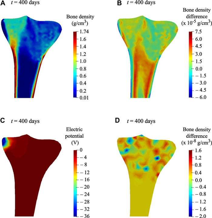
Stress shielding is a critical concept in implantology. If an implant (be it a hip replacement or a dental implant) is significantly stiffer than the surrounding bone, it can carry most of the mechanical load. This "shields" the adjacent bone from the normal physiological strains it needs to maintain its density. According to the mechanostat theory, the local strain falls into the "lazy zone," triggering net bone resorption and potentially leading to loosening of the implant over time.
This is a major consideration in the design of dental implants, influencing their material choice (e.g., titanium's elastic modulus) and geometry, a topic we will explore in detail in the next module.
Conclusion
Today, we have bridged the gap between the mechanical world of forces and the biological world of cellular signaling.
Key Takeaways:
- Wolff's Law states that bone structure adapts to its mechanical environment, gaining mass under load and losing it in disuse.
- This process is driven by mechanotransduction, where osteocytes sense physical forces like fluid shear stress and matrix strain.
- Osteocytes translate these physical cues into biochemical signals—primarily by modulating the RANKL/OPG ratio and sclerostin expression—to direct the activity of osteoclasts and osteoblasts.
- The Mechanostat Theory refines Wolff's Law by proposing specific strain thresholds that trigger bone formation, maintenance, or resorption.
- A critical clinical consequence is stress shielding, where a stiff implant carries too much load, causing adjacent bone to lose density due to insufficient mechanical stimulation.
Preview of the Next Lesson:
We have now examined how bone remodels itself to maintain functional integrity under normal (and abnormal) loading conditions. The next logical step is to understand how bone responds to acute trauma. Our next lesson will cover: Describe the biological stages of bone healing (fracture healing cascade) following a surgical intervention like an osteotomy. This will lay the biological foundation for understanding what happens immediately after an implant site is surgically prepared.
Can't find a good explanation? Sign up and we'll make it for you
Sign up