
Welcome back. In the previous lesson, you traced air from the nose or mouth through the trachea, bronchi, and bronchioles to the alveoli. This lesson explains what makes that air move in the first place.
Breathing, also called ventilation, depends on a chain of linked changes:
- Muscles move the diaphragm and rib cage.
- The size of the thoracic cavity changes.
- Lung volume changes with it.
- Air pressure in the lungs changes.
- Air flows from higher pressure to lower pressure.
By the end, you should be able to explain both inhalation and exhalation as a complete cause-and-effect sequence, rather than as a list to memorise.
The physical idea: changing volume changes pressure
Air is made of rapidly moving gas particles. When those particles occupy a smaller space, they collide with the container walls more frequently, producing a greater pressure. If the same amount of gas has more room, collisions are less frequent and pressure falls.
This is described by Boyle’s law. At a constant temperature, pressure and volume are inversely related:
So:
- if volume increases, pressure decreases
- if volume decreases, pressure increases
Air then moves down a pressure gradient: from a region of higher pressure to one of lower pressure.
For breathing, the two regions to compare are:
- atmospheric pressure: the pressure of air outside the body
- air pressure in the lungs, especially in the alveoli
At the instant when these pressures are equal, there is no net movement of air. To breathe in or out, your body must briefly create a pressure difference.
22.3 The Process of Breathing - Anatomy and Physiology 2e
Read this OpenStax explanation to connect the pressure–volume relationship to the mechanics of ventilation. It gives the physical principle behind every stage of inhalation and exhalation.
In the opening discussion before Figure 22.15, read the Boyle's law explanation. Focus on why expanding a gas-filled space lowers pressure. Then, in the subsection “Pulmonary Ventilation,” read the pressure-gradient explanation. Keep one idea in mind: muscles do not pull air directly; they create a pressure difference that causes air to move.
A useful distinction is that the lungs themselves are not muscular pumps. They are elastic organs that follow the movements of the thoracic cavity, the chest space enclosed by the rib cage and diaphragm. A thin fluid-filled region between the lungs and chest wall helps the lungs stay coupled to the expanding and contracting chest.
The structures that change chest volume
Three components matter most in normal, quiet breathing:
- The diaphragm is a broad sheet of muscle beneath the lungs. When relaxed, it is dome-shaped. When it contracts, it flattens and moves downward.
- The external intercostal muscles lie between the ribs. When they contract, they lift the ribs upward and outward.
- The rib cage protects the lungs but is mobile enough to change the width and depth of the chest.
The thoracic cavity can therefore become larger in two directions:
- vertically, when the diaphragm moves down
- outward and forward, when the ribs rise and move out
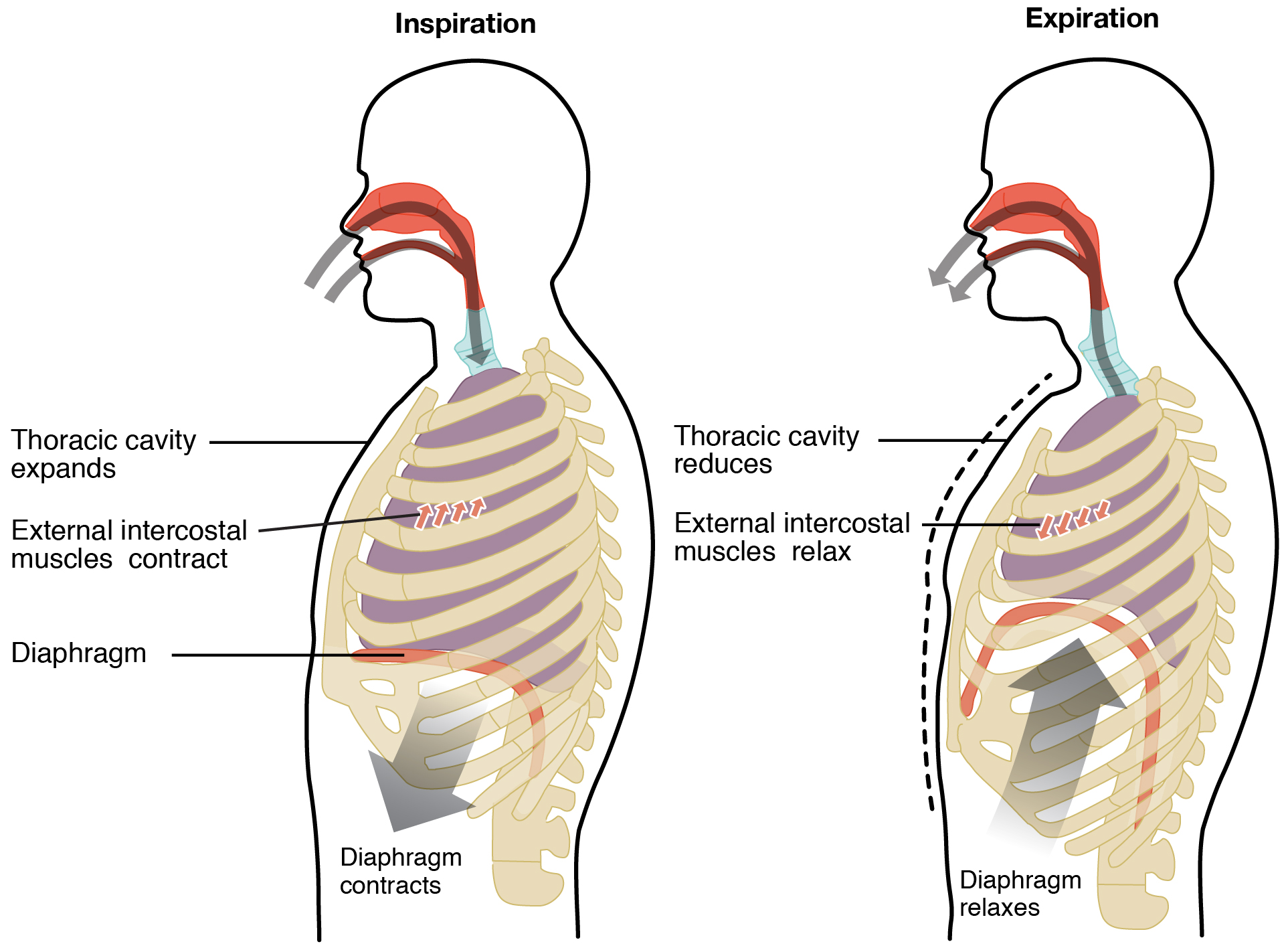
The phrase “the chest expands” is not just a visual description. It is the first physical cause in the sequence that eventually brings air into the lungs.
Ventilation Mechanism | How Breathing Works | Inspiration & Expiration (2026/27 exams)
Watch “Ventilation Mechanism | How Breathing Works | Inspiration & Expiration” from Cognito for a compact visual account of the chest movements and their pressure effects.
Begin with the key structures, identifying the diaphragm, ribs, and intercostal muscles. Then watch pressure and volume to establish the underlying principle. Watch both phases, pausing after inspiration to reconstruct the full chain of events in your own words. Finish with active versus passive for the distinction between quiet and forced exhalation.
Inhalation: making lung pressure lower than atmospheric pressure
Inhalation, also called inspiration, is the process of bringing air into the lungs. It is an active process because muscle contraction requires energy.
During a normal inhalation:
- The diaphragm contracts.
- It moves downward and becomes flatter.
- The external intercostal muscles contract.
- The rib cage moves upward and outward.
- The thoracic cavity increases in volume.
- The lungs are pulled outward and expand.
- The air pressure inside the lungs falls below atmospheric pressure.
- Air flows through the airways into the lungs until the pressures equalise again.
The essential explanation is:
The diaphragm and external intercostal muscles contract, increasing thoracic volume. This increases lung volume and lowers air pressure inside the lungs below atmospheric pressure, so air enters the lungs.
Notice what this statement does not say: it does not say the diaphragm “sucks” air in. Muscles make the chest cavity larger; the resulting low pressure allows outside air to move inward.

You can observe part of this mechanism directly. Place one hand on your lower ribs and another on your abdomen, then take a quiet breath in. The lower ribs move outward and your abdomen moves forward because the contracting diaphragm presses downward into the abdominal region. This outward abdominal movement is an effect of diaphragm contraction, not evidence that air has entered the abdomen.
Exhalation: making lung pressure higher than atmospheric pressure
Exhalation, also called expiration, is the process of moving air out of the lungs.
In normal, quiet breathing, exhalation is usually passive. “Passive” does not mean nothing happens. It means the body does not need to actively contract muscles to push air out during an ordinary breath. Instead, the diaphragm and external intercostal muscles relax, and the elastic lungs and chest wall return toward their resting positions.
During a normal exhalation:
- The diaphragm relaxes.
- It returns upward into a dome shape.
- The external intercostal muscles relax.
- The rib cage moves downward and inward.
- The thoracic cavity decreases in volume.
- The lungs recoil and decrease in volume.
- Air pressure inside the lungs rises above atmospheric pressure.
- Air flows out until the pressures equalise again.
The matching explanation is:
The diaphragm and external intercostal muscles relax, decreasing thoracic volume. This decreases lung volume and raises air pressure inside the lungs above atmospheric pressure, so air leaves the lungs.
The two phases are opposites in terms of volume and pressure, but they are not perfectly symmetrical in energy use:
| Feature | Inhalation | Quiet exhalation |
|---|---|---|
| Diaphragm | Contracts, flattens, moves down | Relaxes, domes upward |
| External intercostals | Contract | Relax |
| Rib movement | Up and out | Down and in |
| Thoracic volume | Increases | Decreases |
| Lung pressure relative to atmosphere | Lower | Higher |
| Direction of airflow | Into lungs | Out of lungs |
| Energy requirement | Active | Usually passive |
Why forced exhalation is different
Quiet exhalation is passive, but a forceful breath out — such as blowing up a balloon, coughing, or exercising intensely — requires more than simple relaxation.
During forced expiration, the internal intercostal muscles can contract to pull the ribs down and inward more strongly. Abdominal muscles may also contract, pushing the diaphragm upward. Both actions reduce thoracic volume further and raise lung pressure more than during quiet exhalation.
This is an extension of the same rule, not a new mechanism:
- a greater decrease in thoracic volume produces a greater rise in pressure
- a greater pressure difference can drive air out more forcefully
For the basic breathing cycle, however, remember that the internal intercostals are not normally needed for a relaxed breath out. The required pair is the diaphragm and the external intercostal muscles.
Reconstructing the whole mechanism
Rather than memorising isolated facts, use this four-link framework whenever you explain ventilation:
| Link | Inhalation | Exhalation |
|---|---|---|
| Muscles and ribs | Diaphragm and external intercostals contract; ribs move up and out | Muscles relax; ribs move down and in |
| Thoracic volume | Increases | Decreases |
| Lung pressure | Falls below atmospheric pressure | Rises above atmospheric pressure |
| Air movement | Air enters | Air leaves |
A common error is to reverse the pressure change. The corrective reasoning is always the same:
- A larger chest produces a larger lung volume.
- Larger volume means particles have more space.
- More space means lower pressure.
- Air therefore enters from the higher-pressure atmosphere.
For exhalation, reverse each part of that reasoning.
Key takeaways
Ventilation is the mechanical movement of air into and out of the lungs. It depends on pressure differences created by changing the volume of the thoracic cavity.
During inhalation, the diaphragm contracts and flattens, while external intercostal muscles raise the ribs upward and outward. Thoracic and lung volume increase, lung pressure falls below atmospheric pressure, and air enters.
During quiet exhalation, these muscles relax. The diaphragm rises, the ribs move down and inward, thoracic and lung volume decrease, lung pressure rises above atmospheric pressure, and air leaves. Quiet exhalation is generally passive because elastic recoil helps reduce lung volume.
Next, you will move from ventilation to gas exchange itself: why the alveoli are structured to make diffusion of oxygen into the blood and carbon dioxide out of the blood fast and efficient.
Can't find a good explanation? Sign up and we'll make it for you
Sign up