
Hello again. In the last lesson, you learned to classify suspicious reconstruction regions as false splits, false merges, missing branches, or uncertain cases. That diagnostic step is only useful if another person—including future you—can return to the exact site and understand why the case was flagged.
This lesson turns an inspection into a compact, auditable candidate-problem note. The aim is not to write a narrative report or to prescribe an edit prematurely. It is to capture enough evidence that the case can be reproduced, reviewed, and acted on safely later.
By the end, you will be able to record four essentials:
- Location — where the candidate is.
- Visual evidence — what you saw in 2D EM and 3D.
- Uncertainty — what is still ambiguous or artifact-limited.
- Proposed next step — the smallest justified follow-up action.
A candidate note is a reproducible bug report
A reconstruction candidate note works much like a good software bug report. “The branch looks wrong” is analogous to “the app is broken”: it may be sincere, but it is not actionable.
A reviewable note lets someone else:
- open the same selected reconstruction;
- navigate to the same coordinates;
- inspect the same local evidence;
- distinguish observation from interpretation;
- know whether the case is ready for review, needs more tracing, or should remain unresolved.
The note is therefore evidence attached to a hypothesis, not a declaration that an edit must be made.
A concise note should answer these questions:
| Field | Question it answers | Good level of detail |
|---|---|---|
| Candidate type | What reconstruction issue is suspected? | False split, false merge, missing branch, or uncertain |
| Location | Where should a reviewer look? | Viewer coordinates plus a nearby recognizable landmark |
| Selected object | Which reconstruction was inspected? | The selected segment/neuron identifier displayed in FlyWire, if available |
| 2D evidence | What does the EM stack show? | Continuity, separate membranes, termination, artifact, and number of sections inspected |
| 3D evidence | What does morphology suggest? | Detached continuation, extra spur, crossing, gap, or other local shape |
| Uncertainty | What prevents a stronger conclusion? | Alternative candidates, fold, misalignment, low contrast, or no unresolved issue |
| Next step | What should happen next? | Review, trace farther, compare candidates, inspect bridge, or leave unchanged |
The minimum viable note is often only three or four sentences. Its strength comes from precision, not length.
The essential distinction: observation, interpretation, decision
The most common note-taking failure is mixing what was seen with what was inferred. Keep these layers separate.
Observation
This is directly visible in the viewer.
Examples:
- “Selected process ends at ; a nearby unselected profile continues for five sections.”
- “Two membrane-bounded profiles remain separate across the apparent crossing.”
- “The section is shifted relative to adjacent images; nearby structures jump together.”
- “The 3D mesh includes a thin spur that turns sharply away from the main arbor.”
Observations should be testable by a reviewer.
Interpretation
This is your classification based on the observations.
Examples:
- “Candidate false split.”
- “Candidate false merge, likely path swap.”
- “Missing branch suspected.”
- “Uncertain because continuity is obscured.”
Use calibrated language: candidate, likely, appears, and supported by are appropriate where evidence is not conclusive.
Decision or next step
This says what remains to be done, without pretending you already completed it.
Examples:
- “Ready for a second reviewer to verify before any edit.”
- “Trace both candidate continuations for an additional 10 sections.”
- “Inspect the suspected bridge at higher magnification and confirm it in 3D.”
- “Leave unresolved; image artifact prevents defensible assignment.”
This separation prevents a premature edit from being smuggled into a note as though it were an observation.
The four-part note format
Use this compact format while inspecting. You can keep it in a plain-text scratchpad, a spreadsheet, a project issue, or an annotation description.
Type:
Location:
Evidence:
Uncertainty:
Next step:
For a higher-value note, add two optional fields:
Selected object:
Reviewer link:
Here is the same template with guidance:
Type: [false split | false merge | missing branch | uncertain]
Selected object: [displayed segment or neuron identifier, if available]
Location: [x, y, z] near [landmark / branch description]

Evidence: In 2D, [what is visible across how many sections].
In 3D, [what the local reconstruction shape shows].

Uncertainty: [none stated / artifact / competing candidates / limitation].
Next step: [bounded review or inspection action].
Reviewer link: [FlyWire shared view, if created]
Two details make this template practical rather than bureaucratic:
- Coordinates alone are not enough. A coordinate identifies a point in a large volume, but a landmark helps a reviewer orient quickly: “thin branch beside dark mitochondrion,” “parallel tract just before image jump,” or “base of the detached twig.”
- Evidence needs a scope. “Several sections” is better than nothing; “five adjacent sections” is better still. It records how much of the stack you actually checked.
Recording a location that another person can find
A location is reproducible when it combines a machine-readable reference with a human-readable one.
1. Capture the viewer coordinates
When you identify the candidate site, record the displayed coordinates exactly as shown in the FlyWire viewer. Do not round them manually. If the suspicious region spans a short bridge or a boundary rather than a single point, place the coordinate near the most diagnostic point:
- for a false split, near the selected termination or the beginning of the validated detached continuation;
- for a false merge, on the suspected erroneous bridge;
- for a missing branch, near the abrupt endpoint or edge of the unexplained 3D gap;
- for an uncertain case, at the artifact or ambiguity boundary.
2. Add a local landmark
Coordinates are efficient for software; landmarks are efficient for people. Add one stable visual cue, such as:
- a mitochondrion-like dark oval;
- a branch bifurcation;
- a conspicuous neighboring large process;
- the beginning or end of a shifted image section;
- a nearby soma or tract, when unambiguous.
Avoid vague descriptions such as “near the top” or “in the middle.” Viewer orientation and zoom change; biological or image landmarks do not.
3. Preserve the selected context
If FlyWire displays an identifier for your currently selected reconstruction, copy it into the note. At this stage, treat it as a reference label, not as a guarantee of stable biological identity; later modules will cover versioned identifiers and edit history.
The Share control can preserve the selected segment and current viewer position as a link. Create a share link after you have centered the evidence location and selected the relevant reconstruction.
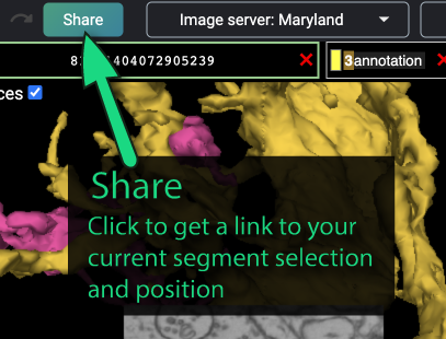
A share link is helpful, but it does not replace coordinates and written evidence. Links can be inaccessible to a collaborator, interfaces change, and a link alone does not state what the reviewer is supposed to verify.
Capturing evidence without overclaiming
The quality of a note depends mainly on its evidence sentence. It should include the relevant facts, but not narrate every mouse movement.
A useful pattern is:
In 2D: [membrane/continuity finding] across [scope].
In 3D: [morphological finding].
Therefore: candidate [classification].
For example:
In 2D, the selected branch terminates immediately before a clear unselected profile; both profiles follow one membrane-bounded process across five adjacent sections. In 3D, the unselected fragment aligns with the branch trajectory. Candidate false split.
Notice what this does not say: it does not say “definitely merge these segments.” The note establishes the evidence needed for later verification.
Evidence appropriate to each classification
| Candidate type | 2D evidence to record | 3D evidence to record |
|---|---|---|
| False split | Where selected material stops and where a separate continuation begins; continuity across adjacent sections | Detached component plausibly extends the selected arbor |
| False merge | Two distinct membrane-bounded processes at the suspected bridge; their separation across slices | Extra spur, X/H structure, abrupt trajectory change, or path swap |
| Missing branch | Endpoint or local region searched; whether a continuation was found | Arbor gap, unusual truncation, or incomplete-looking region |
| Uncertain | Specific artifact, weak boundary, or unresolved alternatives | Whether 3D helps, conflicts, or remains non-diagnostic |
The 2D stack is the primary evidence for physical continuity. The 3D reconstruction is valuable for finding candidates and checking whether an interpretation is morphologically coherent, but a smooth mesh cannot prove that two profiles are biologically connected.
Artifact-aware notes: saying exactly why the case is unresolved
Uncertainty is most useful when it is diagnosed rather than merely asserted. “Uncertain” should be followed by the reason a reviewer cannot make a safe determination.
The FlyWire Proofreading Tips describe how a shifted EM slide, poorly imaged region, or blackout can make the segmentation terminate early or form a poor connection. They also describe using annotation points on a distinctive landmark before and after a jump to compare the displaced views.
Revisit the FlyWire Blog’s practical discussion of image misalignments and difficult thin-neurite continuations. It provides concrete language for distinguishing an actual reconstruction candidate from an image-quality limitation.
In the “Misalignments” section, read from the explanation and annotation workflow. Then continue through “MIA Connections.” Focus on the difference between a continuation you can demonstrate and one that is only plausible after an image jump or disappearance.
Useful uncertainty statements include:
- “A one-section image shift occurs at this location; continuity cannot be evaluated directly across the jump.”
- “Two unselected profiles are plausible continuations after the fold; neither has unique membrane continuity from the pre-artifact branch.”
- “The suspected bridge is visible in the 3D mesh, but 2D inspection has not yet located a definite connection point.”
- “The branch becomes too thin to resolve between closely packed neighboring profiles.”
- “No unresolved limitation found after inspection of six adjacent sections.”
This last form is appropriate when you want to state that the candidate is ready for independent review.
Avoid uncertainty statements that do not explain anything, such as “not sure” or “hard to tell.”
Using annotations as an evidence trail
Annotations can turn your note from a coordinate list into a navigable local case file. The exact interface layout may vary, but the principle is stable: place a small number of purposeful points rather than annotating every slice.
For a difficult site, use up to three annotation points:
- Anchor point — last unquestionably correct location on the selected neurite.
- Candidate point — suspected detached continuation, bridge, or missing endpoint.
- Post-artifact point — a landmark after a misalignment, fold, or blackout.
Name them consistently in your own notes, for example:
A — selected branch before image jump
B — candidate continuation after image jump
C — neighboring landmark used to compare alignment
The annotation panel can list points and export them to CSV, which is useful if you build a small bounded case set instead of a single one-off report.
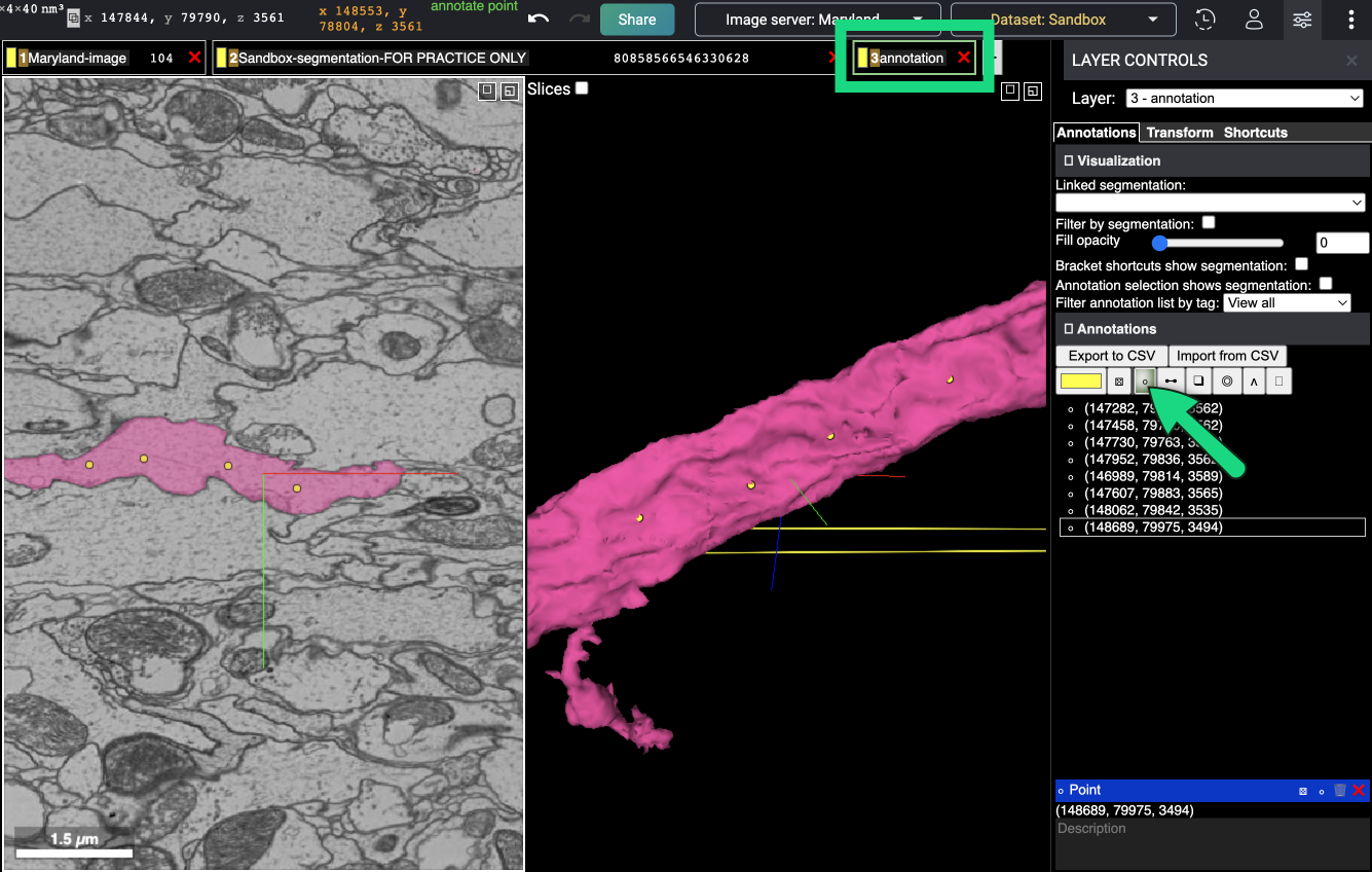
A CSV export stores locations, not your full reasoning. Keep the evidence, uncertainty, and next-step fields in a companion note or structured table. For several cases, a practical local schema is:
| case_id | type | coordinates | landmark | 2D evidence | 3D evidence | uncertainty | next_step | share_link |
|---|
This is intentionally simple. It can later become the basis of an audit log, but for now its purpose is to preserve inspection evidence before any edit is considered.
Worked examples
The following examples are hypothetical, but each follows the same evidence discipline you should use in FlyWire.
Example 1: Well-supported false split
Type: Candidate false split
Selected object: [selected ID shown in viewer]
Location: [x, y, z], thin branch beside a dark oval landmark

Evidence: In 2D, the selected branch ends at the landmark. A separate
nearby segment continues within the same membrane-bounded process for
five adjacent sections. In 3D, that detached segment extends the
trajectory of the selected twig.

Uncertainty: No competing continuation observed in the inspected range.
Next step: Request independent review of the local continuity before
performing any join.
Why this is strong:
- it identifies a specific detached continuation;
- it records a multi-section basis rather than one attractive slice;
- it makes the next step a review, not an unverified action.
Example 2: False merge with a likely path swap
Type: Candidate false merge (possible path swap)
Selected object: [selected ID shown in viewer]
Location: [x, y, z], parallel neurites immediately after image shift

Evidence: In 2D, the selected reconstruction changes from the left
membrane-bounded process to the right process across the shifted section.
The two processes remain distinct in the next four sections. In 3D, the
attached continuation makes an abrupt turn relative to the incoming
branch.

Uncertainty: The true continuation of the original process has not yet
been identified.
Next step: Verify the suspected bridge and trace the original process
beyond the image shift; record any detached true continuation separately.
This note correctly makes the foreign attachment the primary candidate while preserving the possibility that a related false split exists elsewhere.
Example 3: Missing branch, not yet a false split
Type: Missing branch candidate
Selected object: [selected ID shown in viewer]
Location: [x, y, z], distal edge of broad dendritic arbor

Evidence: In 3D, the arbor has a large unfilled wedge and one broad
branch ends abruptly near this location. In 2D, three nearby unselected
segments were inspected, but none was traced with confirmed continuity
to the selected branch.

Uncertainty: Several candidate fragments remain; no specific detached
continuation is validated.
Next step: Search outward from the abrupt endpoint and compare nearby
segments over a wider section range. Do not classify as a false split yet.
The important restraint is the final sentence. A conspicuous gap may be a valuable lead, but it is not proof that a particular object should be added.
Example 4: Correctly unresolved uncertain case
Type: Uncertain
Selected object: [selected ID shown in viewer]
Location: [x, y, z], branch entering a folded/low-quality region

Evidence: The selected thin process is visible before the artifact. Two
profiles appear beyond it, but neither can be connected through visible
membrane continuity. The 3D view does not distinguish the alternatives.

Uncertainty: Folded image region prevents direct tracing; two plausible
continuations remain.
Next step: Preserve anchor and candidate annotations, then leave the
case for targeted review rather than proposing an edit.
This is a successful outcome. The note transforms an ambiguous observation into a bounded, reviewable task.
A fast workflow during live inspection
When you encounter a suspicious region, use this short routine before moving on:
- Freeze the context. Keep the relevant reconstruction selected and center the suspected site.
- Choose the diagnostic location. Record exact viewer coordinates at the termination, bridge, artifact, or candidate continuation.
- Inspect a bounded range. Trace enough adjacent sections to support or limit the claim; record the actual count.
- Check 3D once. Describe only the morphology relevant to the candidate: detached fragment, spur, crossing, gap, or no added clarity.
- State the classification. Use the most specific label your evidence supports.
- Name the uncertainty. If there is none in the inspected range, say so; if present, identify its cause.
- Propose one bounded next step. A reviewer should know exactly what remains to be checked.
- Optionally annotate and share. Add purposeful anchor/candidate points and generate a viewer link for review.
Do not let note-taking turn into a second, unstructured search. If more tracing is necessary, make that tracing the explicit next step and move on.
Quality check before you save the note
Read your note once as though you were a reviewer who has not seen the case. It passes a basic quality check if the answers are clear:
- Can I find it? Exact coordinates, selected context, and landmark are present.
- Can I see why it was flagged? The note names visible 2D and, where useful, 3D evidence.
- Can I tell fact from inference? Observations are distinct from the candidate classification.
- Can I judge confidence? The uncertainty is explicit rather than hidden.
- Can I act responsibly? The next step is bounded and does not demand an unsupported edit.
If any answer is no, improve the note—not the confidence level.
Takeaways and next step
A concise candidate-problem note is the handoff point between inspection and safe proofreading. It should preserve:
- a reproducible location through coordinates, landmark, selected context, and optionally a shared viewer link;
- concrete visual evidence from the EM stack, with 3D morphology used as supporting context;
- explicit uncertainty, especially for folds, shifts, weak boundaries, and competing continuations;
- a narrow next step such as targeted tracing, bridge verification, independent review, or leaving the case unresolved.
Your working standard is simple: a reviewer should be able to reopen the location and understand both what you observed and what you did not establish.
Next module, you will move from documenting candidates to safe edits. You will first learn why segments, supervoxels, and root identifiers determine the scope and consequences of a proofreading action.
Can't find a good explanation? Sign up and we'll make it for you
Sign up