
Hello! Welcome to the first lesson of our fifth module, "Pre-Surgical Assessment and Digital Planning."
In our last lesson, we concluded our deep dive into the biology of osseointegration by analyzing the various local and systemic factors that can lead to implant failure. We saw how issues like insufficient bone volume, micromotion, and proximity to vital structures are significant local risks. The logical next question is: how do clinicians assess and mitigate these risks before ever making an incision?
Today, we address that question directly. Your learning outcome is to explain the role of Cone Beam Computed Tomography (CBCT) in providing 3D anatomical data for implant planning. We will explore what CBCT is, how it works, and most importantly, how it has revolutionized a clinician's ability to "see" the surgical site in three dimensions, transforming implant placement from a procedure based on estimation to one founded on precise digital planning.
From Flat Shadows to 3D Reality
For decades, dental diagnostics relied on 2D imaging like periapical and panoramic radiographs. While useful, these techniques have a fundamental limitation: they compress a three-dimensional structure onto a two-dimensional plane.
This results in:
- Superimposition: Anatomical structures are overlaid, obscuring details.
- Geometric Distortion: The image is not a 1:1 representation of the true anatomy.
- Lack of Cross-sectional Information: Most critically for implantology, the bucco-lingual (cheek-to-tongue) dimension of the jawbone is completely invisible. A ridge that appears wide and robust in a panoramic view might actually be a "knife-edge" with insufficient bone to house an implant.
To understand the evolution from 2D to 3D imaging and the rise of CBCT, the following resource provides a concise history and rationale.
Dental Cone Beam Computed Tomography - StatPearls - NCBI
To start, please read the 'Introduction' section of the StatPearls article titled 'Dental Cone Beam Computed Tomography'. It effectively outlines the limitations of 2D radiography and introduces CBCT as the modern solution for 3D dental imaging.
Please read the 'Introduction' section, which concludes just before the 'Function' heading. Focus on why 2D imaging is limited and how CT, and later CBCT, filled the need for 3D visualization.
How CBCT Works: A Computational Reconstruction
Given your background in physics and numerical simulations, the principle behind computed tomography will be familiar. CBCT is a specialized form of CT designed for the maxillofacial region. Here's how it operates:
-
Acquisition: A cone-shaped X-ray beam is projected through the patient's head onto a 2D flat-panel detector. The X-ray source and detector are mounted on a gantry that rotates around the patient's head (typically 180-360 degrees). In a single rotation, it acquires a series of hundreds of 2D projection images, often called "basis images."
-
Reconstruction: These 2D projections are then fed into a computational algorithm. The most common algorithms are variants of filtered back-projection. The algorithm essentially "reconstructs" the 3D volume that must have existed to produce the acquired set of 2D shadowgrams. The output is a volumetric dataset—a 3D grid of voxels (volumetric pixels), where each voxel has a grayscale value corresponding to the radiodensity of the tissue at that point.
This is fundamentally different from a conventional medical CT scanner, which uses a fan-shaped beam to acquire one thin slice at a time, requiring multiple rotations to build up a volume. The cone-beam approach allows for a much faster scan with a significantly lower radiation dose, making CBCT ideal for in-office dental use.
The 'How Does Cone Beam Computed Tomography (CBCT) Work?' section of the StatPearls article provides a good summary of this process.
The Power of 3D Data: A Clinical Case Study
The true value of this 3D voxel dataset is the ability to explore the anatomy from any angle without distortion. The data can be rendered as a 3D model or, more commonly, sliced into orthogonal planes:
- Axial: Horizontal slices (top-down view).
- Coronal: Vertical slices (front-back view), also called cross-sectional.
- Sagittal: Vertical slices (side-to-side view).
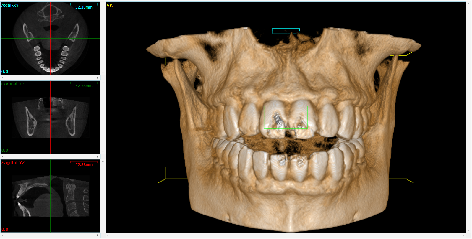
Let's see this in action. The following video is an excellent demonstration of how a clinician uses CBCT to analyze a potential implant site, revealing critical information that was invisible on a 2D radiograph.
In 'Implant #20 CBCT analysis', Dr. Kim masterfully contrasts the ambiguity of a 2D periapical X-ray with the clarity provided by a full CBCT scan for an implant site in the lower jaw.
Please watch from the beginning to 06:10. Observe how he moves through the different views (Axial, Coronal/Cross-sectional, Sagittal) to answer the questions he initially posed about the 2D image. Pay special attention to the discovery of the 'sublingual undercut' in the coronal view—a feature that is impossible to see in 2D and would be catastrophic if not identified before surgery.
As the video vividly illustrates, the CBCT scan transforms planning from guesswork to a precise analysis. The clinician can now answer key questions with certainty:
- What is the true bucco-lingual width of the bone? (Answered by the coronal/cross-sectional view)
- What is the shape of the ridge? Is there a concavity? (The sublingual undercut is a perfect example)
- Where exactly is the mandibular canal? (Traced in 3D)
- What is the available bone height above the nerve? (Measured precisely)
Key Anatomical Data for Implant Planning
The CBCT scan becomes the digital blueprint for the surgery. It is the imaging modality of choice for modern implantology precisely because it provides this comprehensive dataset.
A deep learning approach for dental implant planning in ...
The research paper 'A deep learning approach for dental implant planning' provides a concise summary of the information sought from CBCT scans in its background section.
Read the second paragraph of the 'Background' section (starts with 'CBCT devices...'). It lists the key benefits and applications of CBCT in determining implant size and assessing the need for additional procedures.
Synthesizing from our resources, a clinician interrogates the CBCT data to assess:
- Bone Quantity: Measuring the available bone height, width, and length with sub-millimeter accuracy. This dictates the maximum possible implant dimensions.
- Bone Morphology: Visualizing the 3D shape of the alveolar ridge to identify undercuts, concavities, or knife-edge ridges that could lead to surgical complications like bone perforation.
- Location of Vital Structures: Accurately mapping the 3D path of nerves (e.g., inferior alveolar nerve), the position of the mental foramen, the floor of the maxillary sinus and nasal cavity, and the roots of adjacent teeth. A "safety zone" (typically 2 mm) is planned around these structures.
- Assessment of Pathology: Detecting any underlying issues such as cysts, tumors, retained root fragments, or periapical infections that must be managed before or during implant placement.
The video below gives further examples of identifying the sinus floor and the inferior alveolar nerve in different clinical situations.
#dentalimplant CBCT implant planning and Pano planning
The video '#dentalimplant CBCT implant planning' by Dr. Ivan Chicchon shows more examples of CBCT analysis in both the upper and lower jaw.
Watch the segments from 05:39 to 07:42. The first part shows assessment of bone height relative to the maxillary sinus floor. The second part demonstrates how to scroll through cross-sections to locate the inferior alveolar nerve canal in the mandible.
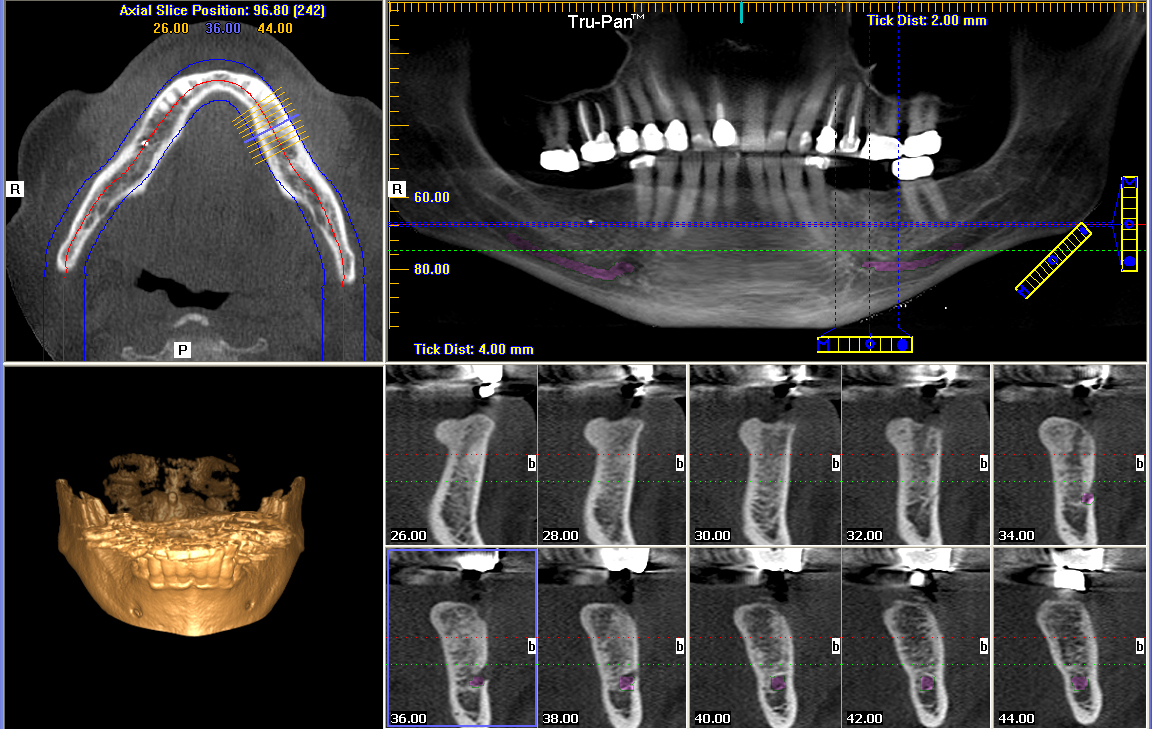
This entire dataset is then displayed in the planning software, allowing the surgeon to virtually place an implant of a specific size and type into the 3D model of the patient's jaw, ensuring it is positioned optimally within the available bone and clear of all critical structures.
Test your understanding!
A clinician is planning an implant in the posterior mandible. A 2D panoramic X-ray shows what appears to be 14mm of bone height above the mandibular canal, which seems sufficient for a 10mm implant. Why is proceeding to surgery based only on this information considered below the standard of care? What specific, critical information is missing that a CBCT would provide?
Show answer
Proceeding based only on the 2D image is risky because it lacks any information about the third dimension (bucco-lingual width). The critical missing information that a CBCT would provide includes:
- The cross-sectional width and shape of the bone. The ridge could be extremely narrow (a "knife-edge"), providing no support for an implant, even if it is tall.
- The presence of lingual concavities. The area below the mylohyoid ridge often has a significant undercut. Drilling based on the 2D image could easily lead to perforation of the lingual cortical plate.
- The precise 3D path of the inferior alveolar nerve. The panoramic view shows a general location, but the CBCT allows for exact tracing of the canal's path, ensuring a safe distance is maintained.
Conclusion
In this lesson, we have established the central role of Cone Beam Computed Tomography in modern dental implantology. By moving from 2D shadows to a 3D volumetric dataset, CBCT provides the essential anatomical information needed for safe and predictable surgical planning.
Key Takeaways:
- Traditional 2D radiographs are insufficient for implant planning because they cannot visualize the cross-sectional dimension of the jawbone.
- CBCT uses a cone-shaped X-ray beam and a computational reconstruction algorithm to create a 3D voxel-based model of the patient's anatomy from a single rotation.
- This 3D data allows clinicians to perform a complete pre-surgical assessment, accurately measuring bone quantity, evaluating bone morphology, and mapping the precise location of critical anatomical structures like nerves and sinuses.
- The role of CBCT is to provide the comprehensive 3D anatomical data required to virtually plan the surgery, mitigating risks and ensuring the implant is placed in an optimal and safe position.
Next Lesson Preview:
We've now seen how CBCT provides the geometric blueprint of the surgical site. However, the quality of the bone is just as important as the quantity. In our next lesson, we will focus on the next step of the assessment: explaining how bone density (measured in Hounsfield units) and morphology are quantified from CBCT scans to assess site suitability. We'll explore the possibilities and limitations of using CBCT data not just for its geometry, but for its physical properties.
Can't find a good explanation? Sign up and we'll make it for you
Sign up