
Hello, and welcome to the Biology revision module on Cells and Biochemical Processes. This module begins with a core exam skill: moving beyond colourful textbook diagrams to identify cell structures in the light and electron micrographs used in questions.
By the end of this lesson, you should be able to identify common organelles from their visual features, state their functions precisely, distinguish plant from animal cells, and use organelle abundance to make sensible inferences about what a specialised cell does.
From diagrams to micrographs
A diagram is simplified: each organelle has a distinctive colour, a clean outline, and a label. A micrograph is an image produced using a microscope. It shows real cells, so structures may be grainy, overlapping, cut at unusual angles, or not visible at all.
There are two broad kinds you need to recognise:
- Light micrographs use visible light. They can show whole cells and larger structures such as cell walls, nuclei, chloroplasts, and large vacuoles. Fine structures such as ribosomes, internal mitochondrial folds, and ER membranes usually cannot be resolved clearly.
- Electron micrographs use electrons rather than light, giving far greater resolution. They usually appear grey, black, and white, but may be false-coloured to make organelles easier to distinguish. They reveal ultrastructure, including membranes, cristae in mitochondria, and ribosomes.
A useful rule is:
Identify an organelle from several pieces of visual evidence, not from one vague shape.
For example, an oval structure is not automatically a mitochondrion. In an electron micrograph, a mitochondrion is more convincingly identified if it has a double boundary and folded internal membranes.
A reliable method for interpreting a micrograph
When a question gives you an unfamiliar cell image, avoid trying to name every organelle immediately. Work from the largest and clearest evidence to the smaller details.
-
Find the cell boundary.
A thick rigid outer layer suggests a plant cell wall. A thin dark boundary alone is likely the plasma membrane. -
Locate the nucleus.
In eukaryotic cells, the nucleus is usually one of the largest, most recognisable structures. It may contain a darker, round nucleolus. -
Decide whether the cell is plant-like or animal-like.
Look for a cell wall, chloroplasts, and a large central vacuole. Their presence is strong evidence for a plant cell. -
Search for distinctive internal patterns.
Mitochondria have folded inner membranes; chloroplasts often show internal stacks; rough ER has ribosomes attached; the Golgi apparatus has curved stacks with small vesicles nearby. -
Use abundance as supporting evidence.
A cell filled with mitochondria probably has a high ATP demand. Extensive rough ER, Golgi stacks and vesicles suggest protein secretion. These clues support an interpretation; they do not replace visual identification.
The following video is a useful visual walkthrough before you begin committing the features to memory.
MICROGRAPHS | The secret to identifying organelles
Watch “MICROGRAPHS | The secret to identifying organelles” by Miss Angler. It demonstrates how the clean organelle shapes in diagrams translate into the grainy, two-dimensional images used in assessments.
Watch plant-cell clues to see how cell walls, membranes, cytoplasm, nuclei, chloroplasts, vacuoles and mitochondria can be recognised in a plant micrograph. Focus particularly on the contrast between chloroplast internal stacks and the curved inner folds of mitochondria. Then watch animal-cell clues for animal-cell boundaries, nuclei, mitochondria, vesicles, endoplasmic reticulum and Golgi apparatus. Notice that ER is an extended membrane network, while the Golgi apparatus is more compact and has nearby vesicles.
The structures you need to recognise and explain
In an exam, a complete response usually has two parts:
- Identification: name the organelle and cite a visible clue.
- Function: state what it does for the cell, using accurate biological language.
| Structure | Recognition clues in a micrograph | Main function |
|---|---|---|
| Plasma membrane | Very thin boundary around the cell; just inside the cell wall in plant cells | Selectively controls movement of substances into and out of the cell |
| Cytoplasm | Granular or lightly textured material filling the cell around organelles | Suspends organelles and is the site of many metabolic reactions |
| Nucleus | Large rounded structure, often with a distinct boundary; may contain a darker nucleolus | Contains DNA and controls cell activities through gene expression |
| Nucleolus | Dense dark circular region inside the nucleus | Produces ribosomal components |
| Ribosomes | Very small dark dots, either free in cytoplasm or attached to rough ER | Assemble amino acids into proteins |
| Rough ER | Parallel or folded membranes with dark dots on the surface; often near the nucleus | Synthesises and transports proteins, particularly those for secretion or membranes |
| Smooth ER | Membranous tubules without attached ribosomes | Synthesises lipids and assists with detoxification and other chemical processing |
| Golgi apparatus | Compact curved stacks of flattened membranes, often with vesicles nearby | Modifies, sorts and packages proteins or lipids for transport |
| Vesicles | Small round membrane-bound sacs, often near Golgi | Transport or store substances |
| Mitochondrion | Oval or elongated shape with a double membrane and internal folds | Produces ATP through cellular respiration |
| Lysosome | Small, round, dense membrane-bound body; difficult to identify with certainty unless context is clear | Contains digestive enzymes that break down materials and worn-out organelles |
| Cell wall | Thick, rigid outer boundary outside the plasma membrane | Supports, protects and maintains the shape of plant cells |
| Chloroplast | Larger oval organelle with visible internal stacks or parallel membranes | Carries out photosynthesis, using light energy to produce glucose |
| Central vacuole | Very large, pale or apparently empty region occupying much of a plant cell | Stores cell sap and maintains turgor pressure, helping support the plant cell |
Two frequent mix-ups are worth fixing now:
- Mitochondria vs chloroplasts: both are oval and have internal membranes. Mitochondrial internal membranes are folded and wavy, called cristae. Chloroplasts have more ordered internal stacks of membranes, called grana.
- Rough ER vs Golgi apparatus: rough ER is usually more extensive and has ribosomes, appearing as small dots on its surface. Golgi stacks are more compact, curved, and associated with budding vesicles.
Why structure and function belong together
An organelle’s structure often makes its function possible. This is more powerful than simply memorising a list.
Take the mitochondrion. Its inner membrane is folded into cristae. These folds create a large surface area for reactions involved in ATP production. Therefore, cells with high energy demands, including muscle cells, commonly contain many mitochondria.
The rough ER is another strong example. Ribosomes attached to its membranes make proteins. A cell that secretes large quantities of protein, such as a pancreatic cell producing digestive enzymes, is likely to have abundant rough ER, a developed Golgi apparatus, and many vesicles for packaging and secretion.
The central vacuole of a plant cell may appear pale because it is largely filled with fluid. When it contains sufficient water, it presses the cytoplasm against the cell wall and helps maintain turgor pressure. This contributes to the firmness of non-woody plant tissues. When a plant loses water, vacuoles shrink and the plant may wilt.
Use the following reading to consolidate the functions behind the visual patterns.
4.3 Eukaryotic Cells - Biology 2e
Read the selected parts of OpenStax Biology 2e’s “Eukaryotic Cells” to connect organelle structures with their functions. Focus on details that explain why particular features, such as a nucleolus or mitochondrial cristae, are visible and biologically significant.
In the “Nucleus,” “Nuclear Envelope,” “Chromatin and Chromosomes,” and “Nucleolus” subsections, read the nucleus section. Track the roles of DNA, the nuclear envelope, nuclear pores and the nucleolus. In the “Ribosomes” and “Mitochondria” subsections, read protein synthesis and ATP production. Pay special attention to why ribosomes appear as dots and why the mitochondrial inner membrane has folds. Finally, in “Animal Cells versus Plant Cells,” read the opening comparison of cell types, then read the chloroplast passage and the central-vacuole passage. Focus on the three high-value plant-cell identifiers: cell wall, chloroplast and central vacuole.
Reading an electron micrograph with confidence
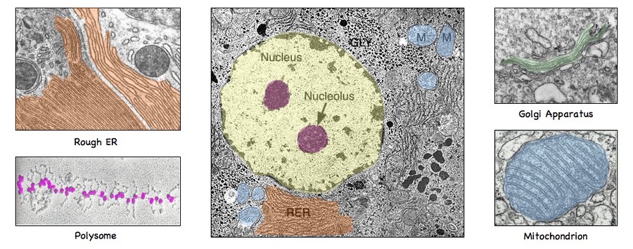
The electron micrograph montage shows why organelles cannot be identified from a single cartoon-like outline:
- The nucleus is large and pale relative to its surroundings, with a darker nucleolus within it.
- Rough ER appears as many closely packed membranes. It is rough because ribosomes are attached to its outer surface.
- A polyribosome is a cluster of ribosomes working on protein production. In a micrograph, ribosomes are tiny dense dots rather than large circular structures.
- The Golgi apparatus is a stack of flattened, curved sacs. Look for small vesicles around the edges.
- A mitochondrion has an outer boundary and internal folds. These cristae are the best identifying feature.
Remember that an electron micrograph is a thin two-dimensional slice through a three-dimensional cell. If a mitochondrion is cut lengthways, it may look elongated; if it is cut across, it may look circular. Therefore, internal membrane structure is more reliable than the overall outline.
Also, darkness is not a direct measure of importance or energy production. It usually reflects how the specimen was prepared and which regions are more electron-dense.
Making functional inferences from organelle abundance
Examiners often ask you to infer the function of a specialised cell rather than merely label its organelles. Use a cautious evidence-based explanation.
A strong structure-function inference has this form:
Observation + organelle function + reasoned conclusion
For example:
The cell contains numerous mitochondria, identified by their folded internal membranes. Mitochondria produce ATP through cellular respiration, so their abundance indicates that the cell has a high energy requirement.
Or:
The cell has extensive rough ER with many attached ribosomes, as well as Golgi stacks and vesicles. These structures are involved in protein synthesis, modification and packaging, indicating that the cell is specialised for producing and secreting proteins.
Common interpretations include:
| Abundant structure | Reasonable inference |
|---|---|
| Many mitochondria | High ATP demand, such as active transport, contraction or sustained cell activity |
| Extensive rough ER and Golgi | Large-scale protein production and secretion |
| Numerous lysosomes | Digestion of engulfed material or recycling of cell components |
| Many chloroplasts | Photosynthesis; likely tissue exposed to light, such as leaf mesophyll |
| Large central vacuole and cell wall | Plant-cell support and water storage |
Be precise with your wording. “Many mitochondria prove this is a muscle cell” is too strong unless the question gives additional evidence. “Many mitochondria indicate a high energy demand” is scientifically justified.
For a short retrieval routine, look again at the electron micrograph montage. Cover its labels, identify the rough ER, nucleus, nucleolus, Golgi apparatus and mitochondrion using visible features, then reveal the labels and correct your reasoning—not just the names.
Key takeaways
- Light micrographs show larger cell features; electron micrographs reveal detailed organelle ultrastructure.
- Identify organelles using multiple visual clues: location, membrane pattern, internal structure and nearby features.
- The nucleus, mitochondria, rough ER, Golgi apparatus, ribosomes, cell wall, chloroplasts and central vacuole are especially important structures to recognise.
- Plant cells are strongly indicated by a cell wall, chloroplasts, and usually a large central vacuole.
- A high-quality exam response links an organelle’s observable structure to its function, then uses that function to justify an inference about the cell.
Next lesson, you will use scale bars and magnification relationships to calculate the actual size of specimens and the magnification of micrographs.
Can't find a good explanation? Sign up and we'll make it for you
Sign up