
Hello again. You now have the foundation: fluid leaves blood capillaries to bathe body cells as tissue fluid; most returns directly to the blood, while the excess enters lymphatic capillaries and becomes lymph. You also identified that lymph carries much more than water, including proteins, lipids, debris, pathogens, and lymphocytes.
This lesson brings those ideas together. The lymphatic system has three central roles:
- maintaining fluid balance in tissues
- supporting immune defence
- absorbing and transporting certain dietary fats
These are normal physiological functions. In professional beauty-therapy language, it is more accurate to describe the system as supporting fluid drainage and immune surveillance than to claim that a treatment “flushes toxins.”
The big picture: a drainage, defence, and transport network
The lymphatic system is a one-way network that begins in body tissues and eventually returns lymph to the bloodstream. Its roles are closely connected because the same fluid that needs draining can also contain foreign material needing immune inspection, while specialised lymphatic capillaries in the intestine transport fats that blood capillaries do not readily take up.
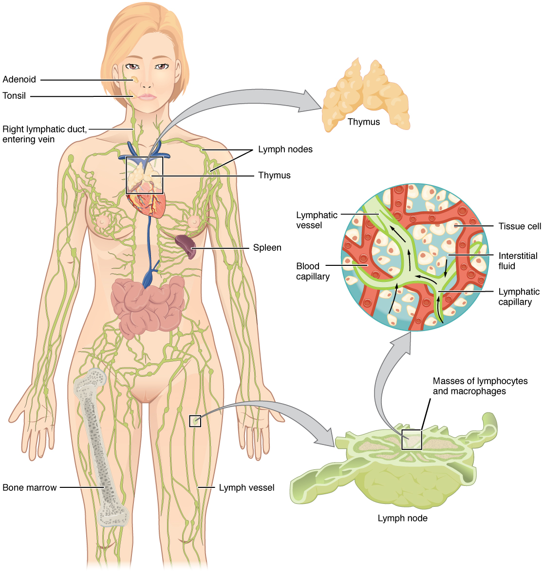
Use the diagram as a map rather than trying to memorise every label now. Notice three useful features:
- The green vessels begin widely throughout tissues, collecting excess tissue fluid.
- Lymph nodes occur along the vessels, placing immune cells in the path of travelling lymph.
- The system returns lymph to the venous circulation near the base of the neck. You will trace this route in detail in the next module.
Watch “Lymphatic System” by Siebert Science for a visual explanation of the three roles before studying each one in more depth.
Begin with three functions for the overall framework. Then watch fluid balance, focusing on the change in name from plasma to tissue fluid to lymph and on why surplus fluid must be collected. Continue with node surveillance to see how lymph is screened for pathogens; the detailed B- and T-cell roles can wait until Module 3. Finally, watch fat absorption, noting the lacteal in an intestinal villus and the route by which dietary fats reach the bloodstream.
1. Fluid balance: preventing persistent tissue swelling
Blood capillaries are exchange sites. Their walls allow fluid and small dissolved substances to move out into the spaces between cells. This fluid delivers oxygen and nutrients to nearby tissues and is called tissue fluid or interstitial fluid.
A substantial proportion of that fluid is reabsorbed into blood capillaries. However, a small excess remains in the tissues. This is not a mistake in the system; it is an expected consequence of capillary exchange. The lymphatic system provides the essential “return route” for that remaining fluid.
At the tissue level, the sequence is:
- Fluid leaves blood capillaries and enters tissue spaces.
- Most is taken back into the blood.
- Excess fluid, along with proteins that have entered the tissue spaces, enters lymphatic capillaries.
- Once inside, it is called lymph.
- The lymphatic system eventually returns it to the bloodstream.
The lymphatic system therefore helps maintain the correct volume of fluid in tissues and helps preserve blood volume. It is particularly important that lymphatic capillaries can collect proteins as well as water. Proteins remaining in tissue spaces tend to draw water towards them, so failure to remove excess protein-rich fluid can make swelling worse.
If lymphatic drainage is impaired by damage, obstruction, disease, or removal of lymphatic structures, fluid can build up in tissues. This is oedema (swelling). A specific form associated with reduced lymphatic drainage is lymphoedema.
It is useful to distinguish two related terms:
| Term | Meaning |
|---|---|
| Oedema | An abnormal accumulation of fluid in tissues; it has many possible causes. |
| Lymphoedema | Persistent swelling associated with impaired lymphatic drainage and often protein-rich tissue fluid. |
In beauty therapy, visible swelling is not automatically evidence of a “slow lymphatic system.” It may have medical, circulatory, inflammatory, hormonal, medication-related, or lymphatic causes. The appropriate response depends on a full consultation and scope of practice, not on a cosmetic assumption.
Introduction to the Lymphatic System - SEER Training Modules
Read this concise overview from the U.S. National Cancer Institute’s SEER Training Modules. It consolidates the three functions using clear terminology that is appropriate for revision.
Start with the first paragraph, which explains excess tissue fluid and proteins. Read the fluid-balance explanation, focusing on why unreturned fluid would contribute to oedema. Then read the next paragraph, beginning “The second function,” through fat absorption. Finish with the short final paragraph, immune defence. Keep the three functions distinct, but notice that all depend on lymph carrying material from tissues.
2. Immune defence: bringing tissue threats to immune checkpoints
The lymphatic system does not merely remove fluid. Because lymph is collected from tissue spaces, it may contain bacteria, viruses, damaged-cell material, or other foreign particles. The lymphatic vessels carry these materials towards lymph nodes.
A lymph node is not simply a storage point, nor is it a place where lymph is made. It is a filtering and immune-response site. As lymph enters a node, it passes slowly through spaces containing immune cells.
The key idea is this:
Lymph nodes allow the body to inspect material collected from tissues before lymph returns to the bloodstream.
Several cell types contribute to this inspection:
- Macrophages can engulf microbes and cellular debris.
- Dendritic cells help alert lymphocytes to foreign material.
- Lymphocytes recognise and coordinate more specific immune responses.
You met lymphocytes in the previous lesson. For this lesson, it is enough to understand that B lymphocytes and T lymphocytes are important immune cells found in lymphoid tissues, including lymph nodes. Their separate roles will be examined later.
The magnified lymph-node inset in Anatomy of the Lymphatic System shows masses of lymphocytes and macrophages inside a node. This illustrates why a node is much more than a swelling along a vessel: it is an active immune checkpoint.
When there is infection or inflammation in a region, nearby lymph nodes may become enlarged or tender. This can happen because immune cells are more active and may multiply in the node. However, swollen nodes also have a range of possible causes. They should never be casually interpreted as a sign that a beauty treatment is “working.”
The lymphatic system therefore contributes to defence in two linked ways:
- it transports pathogens, debris, and immune cells through lymphatic vessels
- lymph nodes filter lymph and provide places where immune responses can begin
The skin is also relevant here. As a physical barrier, it helps prevent pathogens entering the body in the first place. If microbes do enter through damaged skin or an infected area, lymphatic drainage helps bring evidence of that threat to local nodes for immune surveillance.
3. Dietary fats: the intestinal role of lymphatic capillaries
The third role is easy to overlook because it happens in the digestive system rather than in the skin or visible lymph-node regions. Yet it is a major reason the lymphatic system is essential.
The lining of the small intestine has numerous tiny finger-like projections called villi. Villi increase surface area for absorption. Each villus contains blood capillaries, but it also contains a specialised lymphatic capillary called a lacteal.
Most water-soluble nutrients, such as sugars and amino acids, are absorbed into blood capillaries. Many dietary fats follow a different route.
After digestion and processing by intestinal cells, many fats are packaged into particles called chylomicrons. These are too large to enter ordinary blood capillaries easily. Instead, they enter the more permeable lacteals at the centre of intestinal villi.

The healthy upper section of the diagram shows the main idea:
- Dietary fat is processed by intestinal cells.
- Fat is packaged into chylomicrons.
- Chylomicrons enter the lacteal.
- They travel in lymph through intestinal lymphatic vessels.
- The lymph eventually enters venous blood circulation.
Lymph leaving the intestine after a fatty meal can look milky because of its high fat content. This lipid-rich lymph is called chyle.
A useful terminology set is:
| Term | Meaning |
|---|---|
| Lacteal | A specialised lymphatic capillary in an intestinal villus |
| Chylomicron | A particle that packages dietary fats for transport |
| Chyle | Milky, lipid-rich lymph from the intestine |
| Fat-soluble vitamins | Vitamins that can travel with dietary fats; these include vitamins A, D, E, and K |
Be precise with the claim that “fats cannot enter the blood.” The clearer statement is:
Many long-chain dietary fats are packaged into chylomicrons and enter intestinal lacteals before reaching the bloodstream through the lymphatic system.
This route is not a secondary or optional detour. It is a normal part of dietary-fat transport.
Connecting the three roles
Although fluid balance, immune defence, and fat absorption may seem separate, they depend on the same core design: lymphatic capillaries collect material from tissues, lymphatic vessels transport it, and lymph eventually returns to the blood.
| Role | What the lymphatic system collects or carries | Why it matters |
|---|---|---|
| Fluid balance | Excess tissue fluid and proteins | Limits fluid accumulation and helps maintain tissue and blood-fluid volumes |
| Immune defence | Pathogens, debris, immune cells, and tissue-derived information | Lymph nodes filter lymph and support immune responses |
| Dietary-fat absorption | Chylomicrons containing dietary fats and fat-soluble vitamins | Enables fats from the small intestine to reach venous circulation |
For your professional understanding, the important conclusion is not that every appearance of puffiness is a lymphatic problem. Rather, the lymphatic system normally has an ongoing role in collecting excess tissue fluid, returning it to blood, and supporting immune monitoring. When a client has signs of acute infection, unexplained swelling, or a diagnosed lymphatic condition, treatment decisions require appropriate caution, consultation, and sometimes medical referral.
Key takeaways
- The lymphatic system maintains fluid balance by collecting excess tissue fluid and proteins and returning them to the bloodstream.
- If lymphatic drainage is impaired, protein-rich fluid may accumulate, contributing to lymphoedema.
- Lymphatic vessels transport material from tissue spaces to lymph nodes, where macrophages, dendritic cells, and lymphocytes support immune defence.
- Lymph nodes filter lymph; the spleen, by contrast, primarily filters blood.
- In the small intestine, lacteals absorb many dietary fats packaged as chylomicrons.
- Lipid-rich intestinal lymph is called chyle, and it also transports fat-soluble vitamins.
- Professional terminology should focus on normal fluid drainage, transport, and immune surveillance—not vague claims about “detoxing.”
Next, you will follow lymph’s physical route through the body: from lymphatic capillaries to larger vessels, lymph nodes, trunks, ducts, and finally the veins near the neck.
Can't find a good explanation? Sign up and we'll make it for you
Sign up