
Hello! Welcome back to your course on Medical Instrumentation.
In our last module, we focused on sensing and interpreting physiological and behavioral signals from the body's surface, which is highly relevant to wearable technology. We concluded by discussing how context-aware computing is vital for adding meaning to that data.
Now, we shift our focus from sensing on the body to looking inside it with powerful clinical imaging technologies. This lesson addresses the learning outcome: Explain the basic principles of X-ray, Computed Tomography (CT), and Magnetic Resonance Imaging (MRI).
We will explore how each of these revolutionary techniques works, what kind of information it provides, and its fundamental trade-offs. This knowledge is crucial for your exam preparation and provides a foundational understanding of the gold-standard tools used in clinical diagnostics and neuroscience research.
To start, let's look at how these three modalities visualize the same object—a human hand.
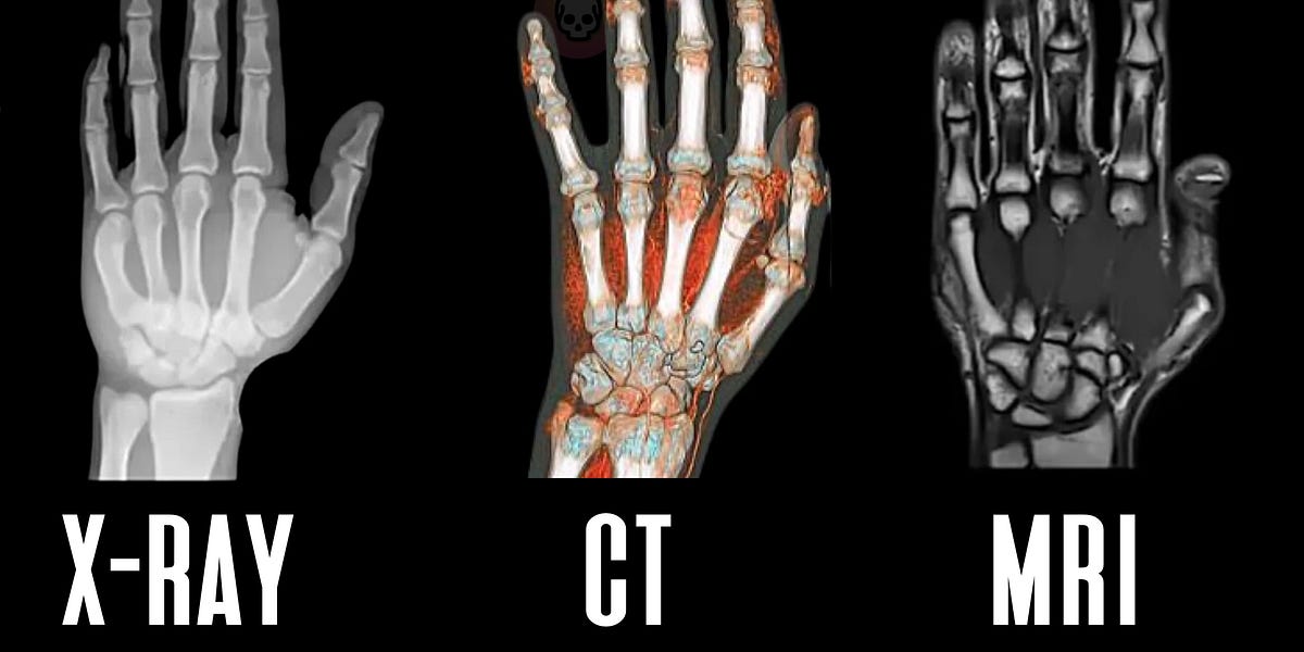
This visual comparison sets the stage for our exploration. Let's break down the principles behind each one.
1. X-ray Imaging: The "Shadowgram"
The oldest and most common form of medical imaging is based on a simple principle: differential attenuation.
When X-rays—a form of high-energy electromagnetic radiation—pass through the body, they are absorbed or scattered to different degrees by different tissues. This process is called attenuation. The amount of attenuation depends on the tissue's density and atomic number.
The basic physics can be described by the following equation:
Where:
- is the initial intensity of the X-ray beam.
- is the intensity after passing through a thickness of tissue.
- is the linear attenuation coefficient of the tissue.
Dense tissues like bone have a high , absorbing many X-rays. Less dense tissues like fat, muscle, and air have a low , allowing more X-rays to pass through. The X-rays that exit the body strike a detector (or photographic film), creating a 2D projection image—essentially a shadow.
Imaging and detectors for medical physics Lecture 3: X-ray ...
These slides from a lecture on X-ray imaging provide a concise overview of the core principles and instrumentation.
Please review the following slides: "X-ray in the body" and "X-ray transmission imaging" (Slides 4-5): Focus on the attenuation equation and the concept of differential absorption. "X-ray imaging techniques" (Slide 7): Note the definition of planar radiography (a 2D projection). "CT –vs– planar radiography" (Slide 49): Read the table comparing the two. This summarizes the key advantages and disadvantages of standard X-ray.
In summary, a conventional X-ray:
- Principle: Differential attenuation of X-rays by body tissues.
- Output: A 2D projection image.
- Strengths: Fast, inexpensive, excellent for imaging dense structures like bones and teeth.
- Weaknesses: Uses ionizing radiation, provides poor contrast for soft tissues, and collapses 3D anatomy into a 2D image, which can obscure structures.
2. Computed Tomography (CT): Slicing with X-rays
Computed Tomography (CT) was developed to overcome the limitations of conventional X-rays, particularly the poor soft-tissue contrast and 2D projection. The core idea is to use X-rays to create cross-sectional "slices" of the body, which can then be stacked together to form a 3D image.

A CT scanner acquires hundreds of X-ray projections from different angles as the gantry rotates around the patient. A powerful computer then processes this massive amount of data to reconstruct a detailed cross-sectional image.
To understand why so many angles are needed, let's watch a video that uses a brilliant analogy.
CT physics overview | Computed Tomography Physics Course | Radiology Physics Course Lesson #1
This video from Radiology Tutorials provides an excellent overview of CT physics. We'll focus on a key section that intuitively explains the need for multi-angle projections.
Please watch the segment from 11:20 to 18:20. The video uses a Sudoku puzzle as an analogy to demonstrate why you cannot reconstruct a detailed image from just one or two projection angles and why the 'computed' part of CT is so essential. This is a very powerful and intuitive explanation.
Image Reconstruction and Hounsfield Units
The mathematical process of creating an image from the projection data is called image reconstruction. A common algorithm for this is filtered backprojection, a concept from signal processing that you may have encountered in your ECE studies.
The result of reconstruction is a grid of pixels, where each pixel's brightness corresponds to the average attenuation of the tissue in that specific volume element (or voxel). These attenuation values are standardized on a scale called Hounsfield Units (HU).
- Air: -1000 HU (very low attenuation)
- Water: 0 HU (the reference point)
- Bone: +1000 HU or higher (very high attenuation)
Clinicians can then use a technique called windowing to adjust the range of HU values displayed in grayscale, allowing them to optimize the image contrast for viewing specific tissues, like bone, lung, or soft tissue.
Imaging and detectors for medical physics Lecture 3: X-ray ...
Let's return to the lecture slides to formalize these concepts.
Please review the following slides: "Computed Tomography (CT)" (Slide 21): Reinforces the basic principle of the rotating source and detector. "2D image reconstruction in CT" (Slide 26): Briefly look over the steps involved in reconstruction. "CT number" and "2D image display" (Slides 34-36): Focus on the definition of the Hounsfield unit and the concept of windowing.
In summary, a CT scan:
- Principle: X-ray attenuation measurements taken from many angles, followed by computational reconstruction.
- Output: Cross-sectional (tomographic) images that can be viewed as a 3D volume.
- Strengths: Fast, provides excellent anatomical detail and good spatial resolution, and eliminates the overlap of structures seen in 2D X-rays.
- Weaknesses: Involves a significantly higher dose of ionizing radiation than a single X-ray, is more expensive, and can still have limited soft-tissue contrast compared to MRI.
3. Magnetic Resonance Imaging (MRI): Listening to Protons
MRI is fundamentally different from X-ray and CT. It does not use ionizing radiation. Instead, it uses a powerful magnetic field and radio waves to generate images. This makes it exceptionally safe and ideal for imaging sensitive areas like the brain, as well as for repeated scans.
The physics behind MRI is complex, but the core principles rely on the behavior of hydrogen protons (the nucleus of a hydrogen atom) in the body. Since our bodies are mostly water and fat, we have an abundance of hydrogen.
The entire process can be broken down into a few key steps. The following video provides a fantastic and engaging explanation of this intricate process.
The Insane Engineering of MRI Machines
The YouTube channel Real Engineering has produced a detailed animation explaining the complex physics and engineering of MRI machines in an accessible way. Watching this is the best way to grasp the core concepts.
Please watch the video from 01:47 to 13:13. This is the main activity for this section. Focus on understanding these sequential steps: Alignment (01:47 - 02:55): How the main magnetic field makes hydrogen protons align. Excitation & Signal (02:55 - 05:18): How a radiofrequency (RF) pulse 'nudges' the protons and how a signal is generated as they 'relax'. Spatial Localization (09:08 - 10:47): How 'gradient magnets' are used to create slices and pinpoint where the signal is coming from. Tissue Contrast (10:47 - 13:13): The crucial concepts of T1 and T2 relaxation and how they allow MRI to differentiate between different types of soft tissue.
Key MRI Concepts Recap
As the video explained, the magic of MRI lies in these concepts:
- Alignment: A strong magnetic field () causes a slight majority of hydrogen protons to align with it.
- Excitation: A radiofrequency (RF) pulse, transmitted at a specific frequency called the Larmor frequency, knocks these aligned protons out of alignment.
- Relaxation: When the RF pulse is turned off, the protons relax back to their aligned state. As they do, they release the absorbed energy as a faint radio signal, which is detected by receiver coils.
- Contrast (T1 and T2): The key to MRI's incredible soft-tissue detail is that protons in different tissues relax at different rates.
- T1 relaxation (spin-lattice): The time it takes for protons to realign with the main magnetic field. T1-weighted images provide excellent anatomical detail.
- T2 relaxation (spin-spin): The time it takes for the processing protons to lose phase coherence with each other. T2-weighted images are excellent for highlighting pathology (like tumors or inflammation) because these tissues tend to have high water content and thus a long T2 time, making them appear bright.
- Image Reconstruction: Similar to CT, a computer is needed to reconstruct the final image. The detected signals are localized in space using magnetic field gradients, and a mathematical technique you're familiar with from your ECE background, the Fourier Transform, is used to convert the raw signal data (in the frequency domain) into a spatial image.
For your startup's focus on neurotechnology, it's worth noting that MRI is a cornerstone of neuroscience. A special type called functional MRI (fMRI) detects changes in blood flow associated with brain activity, allowing researchers to see which parts of the brain are active during specific tasks or emotional states. While not a wearable technology, fMRI provides the foundational brain maps that guide much of the research in your field.
Test your understanding!
For each of the following clinical scenarios, which imaging modality (X-ray, CT, or MRI) would be the most appropriate first choice, and why?
- A patient comes to the emergency room with a suspected broken arm after a fall.
- A neurologist wants to investigate a patient for a suspected brain tumor, which is a soft tissue mass.
- A doctor needs to get a quick, detailed look at a patient's lungs and abdomen after a major car accident to check for internal injuries.
Show answer
- X-ray: It is fast, inexpensive, and provides excellent images of bones, making it perfect for quickly diagnosing a fracture. The radiation dose is low.
- MRI: It is the best choice because it does not use ionizing radiation (critical for the brain) and provides superior soft-tissue contrast, which is necessary to visualize a tumor against surrounding healthy brain tissue. T2-weighted images would be particularly useful.
- CT: It is much faster than an MRI, which is crucial in a trauma situation. It provides excellent cross-sectional views that can quickly identify internal bleeding, organ damage, and complex fractures throughout the abdomen and chest, giving a much more comprehensive view than a simple X-ray.
Conclusion
In this lesson, we've covered the fundamental principles of three landmark medical imaging technologies.
Key Takeaways:
- X-ray works by differential attenuation, creating a 2D "shadow" image that is excellent for visualizing dense structures like bone but poor for soft tissue.
- CT improves on X-ray by using a rotating source and detector to take many projections, which are then computationally reconstructed into detailed 3D images. Its main drawback is a higher dose of ionizing radiation.
- MRI uses powerful magnetic fields and radio waves to manipulate hydrogen protons. It does not use ionizing radiation and provides unparalleled soft-tissue contrast by exploiting differences in tissue relaxation times (T1 and T2).
Preview of the Next Lesson:
Having looked at imaging with electromagnetic radiation (X-ray/CT) and magnetic fields (MRI), we will next explore a modality that uses a completely different physical principle: sound. In our next lesson, we will learn how diagnostic ultrasound uses high-frequency sound waves to create real-time images of soft tissues and blood flow, another cornerstone of non-invasive diagnostics.
Can't find a good explanation? Sign up and we'll make it for you
Sign up