
Hello, and welcome to the course. Over the next eight weeks, you will build a connected picture of the human body: how its systems move materials, exchange gases, control activity, create movement, and keep internal conditions stable. We begin with the body’s levels of organization, because every later topic—from neurons to kidneys—depends on knowing what kind of biological structure we are discussing.
This first module focuses on cells, how they are organized, and how the body maintains relatively stable internal conditions. In this lesson, you will learn to distinguish a cell, tissue, organ, and organ system using both definitions and a practical classification method.
The body is organized in nested levels
The human body is not simply a collection of parts placed next to one another. It is a hierarchy: small living units join together to make more complex structures, and those structures cooperate at progressively larger scales.
Organization in the human body | Cells and organisms | Middle school biology | Khan Academy
Watch “Organization in the human body” from Khan Academy for a compact visual introduction to the four levels. It establishes the core definitions and uses the respiratory system to show how the levels fit together.
Watch the overview for the four-level hierarchy. Continue with cells and tissues, then organs and systems. Finish with the recap, which summarizes the nested relationship. As you watch, focus on one decisive distinction: a tissue is built from similar cells, whereas an organ combines different tissue types.
The four levels relevant to this course are:
- Cell: the smallest unit that is alive and can perform life processes.
- Tissue: a group of similar, specialized cells working together on a shared task.
- Organ: a structure made of two or more tissue types working together for a specific function.
- Organ system: several organs cooperating to carry out a major body function.
A complete person is an organism, containing all of these levels. The hierarchy is nested: the material that makes up one level also contributes to the next.
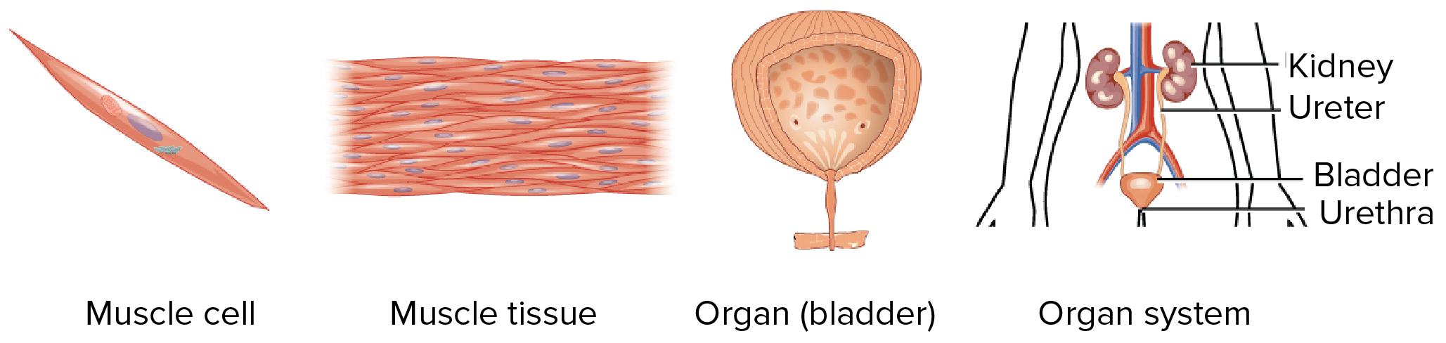
The image is useful because it shows that the labels do not merely reflect size. A bladder is larger than a patch of muscle tissue, but it is called an organ specifically because it contains several tissue types coordinated for urine storage and release.
Level 1: Cells are specialized living units
A cell is the smallest structure that can be considered alive. Cells take in materials, use energy, respond to their surroundings, and maintain internal conditions. Human cells are specialized: their structures fit the jobs they perform.
For classification, names ending in “cell” are usually straightforward:
- Neuron or nerve cell: a cell specialized for receiving and transmitting information.
- Red blood cell: a cell specialized for transporting oxygen.
- Epithelial cell: a cell that helps cover or line a surface.
- Smooth muscle cell: a cell capable of contracting.
- Sperm cell and egg cell: reproductive cells.
A cell can be remarkably elaborate, but it is still one individual living unit. A single muscle cell is therefore a cell, not muscle tissue.
A useful question is:
Am I looking at one individual specialized living unit, rather than a collection of units?
If yes, classify it as a cell.
Level 2: Tissues are coordinated collections of similar cells
A tissue is a group of similar cells, often along with the materials around them, working together on a particular task. Tissue is not just “a lot of cells.” The cells must be organized and function as a group.
For example, a single epithelial cell helps form a lining. A sheet of many epithelial cells lining the mouth, skin, or intestine is epithelial tissue. Its tightly packed arrangement makes it effective as a protective barrier.
The human body has four broad tissue categories:
| Tissue type | Main role | Familiar examples |
|---|---|---|
| Epithelial tissue | Covers surfaces, lines spaces, forms barriers and glands | Outer skin layer; intestinal lining |
| Connective tissue | Supports, connects, stores, or transports | Bone, cartilage, blood, tendons |
| Muscle tissue | Contracts to produce movement | Skeletal, cardiac, and smooth muscle tissue |
| Nervous tissue | Detects, processes, and transmits information | Brain, spinal cord, peripheral nerves |
The category names can help, but function and composition are more reliable than vocabulary alone. Consider blood. It is not usually described as a “solid body part,” but it is classified as a specialized connective tissue: its cells are suspended in a liquid extracellular matrix called plasma, and it transports materials through the body.
Similarly:
- A neuron is a cell.
- Nervous tissue is many neurons and supporting cells organized for signaling.
- Cardiac muscle tissue is a tissue.
- A named structure made partly of muscle tissue, blood vessels, nerves, and connective tissue may be an organ.
The key question is:
Is this mainly a collection of similar cells performing one shared kind of work?
If yes, it is a tissue.
Level 3: Organs combine different tissues for a larger job
An organ contains at least two tissue types arranged so that their different abilities produce one coordinated function. Organs are not just “bigger tissues.” Their defining feature is tissue diversity working together.
Take the small intestine. It has:
- epithelial tissue at its inner surface to absorb nutrients and release some substances;
- smooth muscle tissue to mix and propel food;
- connective tissue to support its structure and carry blood vessels;
- nervous tissue to coordinate local activity.
Together, these tissues allow the small intestine to process food and absorb nutrients. That coordinated function makes it an organ.
The heart provides another clear example. It includes:
- cardiac muscle tissue, which contracts to pump blood;
- connective tissue, which supports and helps organize the heart’s structures;
- nervous tissue, which participates in coordinating contractions;
- epithelial tissue lining surfaces and blood vessels.
The heart is therefore an organ, not “cardiac muscle,” even though cardiac muscle is central to its function.
Common organs include the:
- heart
- lungs
- brain
- skin
- stomach
- liver
- kidneys
- urinary bladder
- small intestine
A helpful diagnostic question is:
Does this structure contain multiple tissue types that cooperate to carry out one recognizable bodily function?
If yes, it is an organ.
A naming trap: “muscle” can mean two different levels
Everyday language can be imprecise. “Muscle” might mean muscle tissue, but it can also mean a named skeletal muscle such as the biceps brachii.
- Skeletal muscle tissue is a tissue: it consists mainly of similar contractile muscle cells.
- The biceps brachii muscle is an organ: it contains muscle tissue plus connective tissue, blood vessels, and nerves.
When a question gives a general tissue label, such as “smooth muscle tissue,” classify it as a tissue. When it names an anatomical structure, such as “the biceps” or “the diaphragm,” classify it as an organ.
Level 4: Organ systems coordinate several organs
An organ system is a group of organs that work together to accomplish a major function that no single organ could fully perform on its own.
The urinary system, shown in the image, includes the kidneys, ureters, bladder, and urethra. The kidneys filter blood and form urine; the ureters carry urine; the bladder stores it; and the urethra provides the route out of the body. Each organ has a distinct contribution, but the coordinated set is one organ system.
Other examples include:
| Organ system | Some components | Broad function |
|---|---|---|
| Respiratory system | Nose, trachea, lungs, diaphragm | Brings in oxygen and removes carbon dioxide |
| Cardiovascular system | Heart, blood vessels, blood | Moves blood, gases, nutrients, hormones, and wastes |
| Digestive system | Mouth, stomach, intestines, liver, pancreas | Breaks down food and absorbs usable materials |
| Nervous system | Brain, spinal cord, nerves, sensory organs | Detects information and coordinates rapid responses |
| Musculoskeletal system | Bones, joints, skeletal muscles | Supports the body and enables movement |
| Endocrine system | Hormone-producing glands | Coordinates longer-lasting changes through hormones |
Systems are useful categories, but they are not sealed off from one another. For example, the respiratory system brings oxygen into the body, while the cardiovascular system transports that oxygen to cells throughout the body. The digestive system supplies nutrients, and the urinary system helps remove certain wastes and regulate water and salt balance.
This interdependence is the central theme of the course: body systems have distinct roles, but survival depends on their cooperation.
A reliable method for classification
When you encounter an unfamiliar example, do not classify it based only on whether it “sounds small” or “sounds important.” Instead, use evidence about what it is made of and how it functions.
Step 1: Look for an individual cell
If the example is one named cellular unit, it is a cell.
- red blood cell
- neuron
- epithelial cell
- sperm cell
Step 2: Look for a group of similar cells
If it is a group of similar specialized cells performing a shared job, it is a tissue.
- muscle tissue
- epithelial tissue
- nervous tissue
- bone tissue
- blood
Step 3: Look for multiple tissue types in one structure
If it contains different tissues integrated for a specific task, it is an organ.
- heart
- kidney
- lung
- skin
- stomach
- bladder
Step 4: Look for several organs cooperating
If the example names a coordinated group of organs with a broad body function, it is an organ system.
- digestive system
- respiratory system
- urinary system
- cardiovascular system
Here is the same approach as a compact decision guide:
| Clue in the description | Likely classification |
|---|---|
| “One specialized living unit” | Cell |
| “Similar cells working together” | Tissue |
| “Several tissue types with one specific function” | Organ |
| “Several organs with a major shared function” | Organ system |
Worked classifications
Let’s apply the method carefully.
Example 1: Red blood cell
A red blood cell is one specialized cell that carries oxygen. It is a cell.
The fact that millions circulate in the bloodstream does not change the classification of one red blood cell.
Example 2: The lining of the small intestine
This is a layer of similar epithelial cells specialized for absorption and barrier functions. It is epithelial tissue.
It is not the entire small intestine. The full small intestine contains several tissues, so it is an organ.
Example 3: The kidney
A kidney contains epithelial tissue in its filtering tubes, connective tissue and blood vessels, nervous tissue, and muscle tissue in associated structures. It performs the integrated job of filtering blood and regulating water and solute balance. It is an organ.
Example 4: The respiratory system
The respiratory system includes the nose, airways, lungs, and diaphragm. These organs collaborate to move air and enable gas exchange. It is an organ system.
Example 5: Smooth muscle
This depends on the wording:
- Smooth muscle tissue is a tissue.
- The smooth-muscle-rich wall of the bladder is only one tissue component of an organ.
- The urinary system, which includes the bladder and kidneys, is an organ system.
The distinction is not a matter of memorizing labels. It follows from composition: similar cells form tissue; multiple tissue types form an organ; multiple organs form a system.
Why organization matters for the rest of the course
Each level gives us a different explanatory lens. If we ask why a neuron can transmit a signal, we are studying a cell. If we ask how a muscle contracts as a coordinated group, we are studying tissue. If we ask how the heart pumps blood, we are studying an organ. If we ask how oxygen reaches a working leg muscle, we need several organ systems at once.
The same biological event can therefore be described at multiple levels. During exercise:
- muscle cells use energy;
- muscle tissue contracts;
- skeletal muscles act through joints to move the body;
- the cardiovascular and respiratory systems deliver oxygen and remove carbon dioxide;
- the nervous and endocrine systems coordinate the response.
Keeping the levels distinct prevents a common confusion: an organ system does not replace its organs, and an organ does not replace its tissues. Each level explains a different part of the whole-body story.
Key takeaways
- A cell is one specialized living unit, such as a neuron or red blood cell.
- A tissue is a group of similar cells working together, such as epithelial, muscle, connective, or nervous tissue.
- An organ contains two or more tissue types coordinated for a particular function, such as the heart, lung, kidney, skin, or bladder.
- An organ system is a group of organs with a major shared function, such as the respiratory, digestive, or urinary system.
- For difficult cases, classify by composition and coordinated function, not just by the everyday name of the structure.
Next, we will move down to the cell level and examine how substances cross the cell membrane. That process of selective movement is essential for everything from nutrient absorption to gas exchange and nerve signaling.
Can't find a good explanation? Sign up and we'll make it for you
Sign up