
Hello! Welcome to the first lesson in your course on dental implantation.
Given your background in physics and research, you're accustomed to building complex models from first principles. We'll take a similar approach here. Before we can understand the dynamics of placing an implant and how it integrates with the body, we first need a precise map of the environment.
This lesson addresses the first learning outcome: Identify the major bones of the maxilla and mandible and their key structural features relevant to implantation. We will systematically explore the anatomy of the upper jaw (maxilla) and lower jaw (mandible), focusing on the specific bony landmarks, cavities, and passages for nerves and blood vessels that a surgeon must navigate. This foundational knowledge is the bedrock upon which all subsequent topics—from surgical planning to biomechanics—are built.
Let's begin by examining the upper jaw.
1. The Maxilla: The Upper Jaw's Complex Architecture
The maxilla is not a single bone but a pair of bones (left and right) that fuse at the midline. It forms the upper jaw, supports the upper teeth, and contributes to the structure of the nasal cavity and the eye sockets (orbits). Its complex, relatively delicate structure presents unique challenges for dental implantation.
To start, let's get a clear visual of its structure.
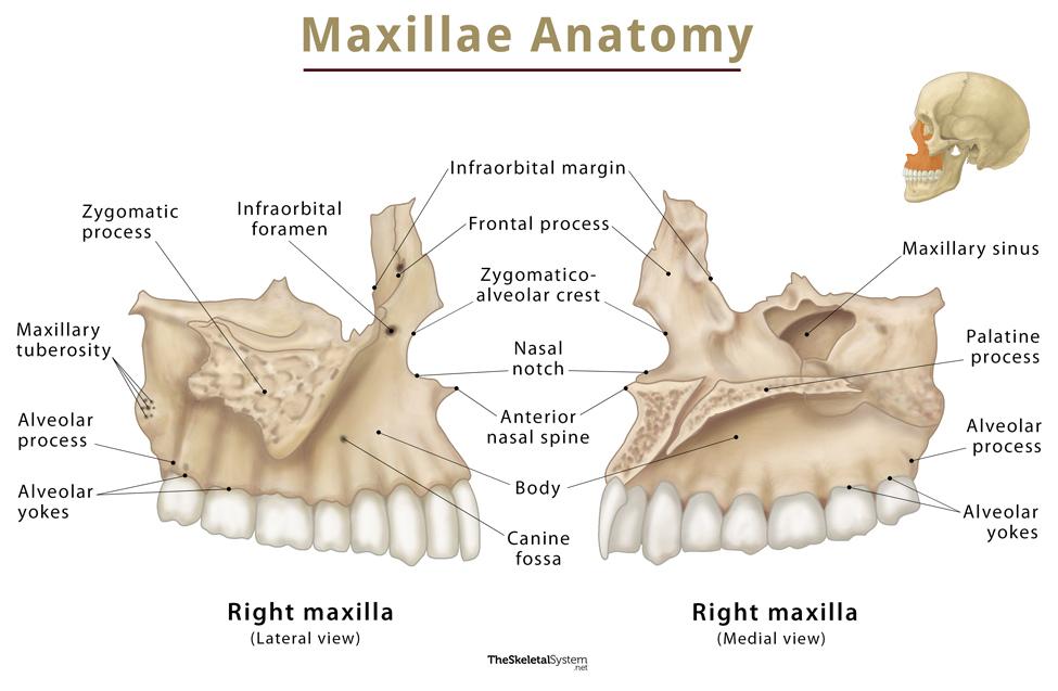
Now, for a detailed, text-based description, please read the following sections from a StatPearls article, a peer-reviewed resource from the National Center for Biotechnology Information (NCBI). This will provide a comprehensive anatomical foundation.
Anatomy, Head and Neck, Maxilla - StatPearls - NCBI - NIH
This reading from 'Anatomy, Head and Neck, Maxilla' on StatPearls provides a detailed, encyclopedic overview of the maxillary bone, its processes, and associated structures.
Please read the 'Introduction' and the 'Structure and Function' sections. Focus on understanding the four main processes (alveolar, palatine, zygomatic, frontal) and the overall pyramidal shape of the maxillary body.
Key Maxillary Features for Implantation
As you've just read, the maxilla is more than just a bone for holding teeth. Let's distill the information and highlight what is most critical from an implantology perspective.
-
Alveolar Process: This is the horseshoe-shaped ridge of bone that contains the tooth sockets (alveoli). When a tooth is lost, this is the bone that will receive the implant. Its height and width are primary determinants of whether an implant can be placed without additional bone grafting.
-
Maxillary Sinus: This is arguably the most significant feature for implants in the posterior (back) maxilla. It is a large, air-filled cavity within the body of the maxilla. The floor of the sinus is located just above the roots of the upper premolars and molars. After tooth extraction, the sinus often expands downwards (a process called pneumatization), reducing the available bone height for an implant.
-
Incisive Canal (or Nasopalatine Canal): Located in the anterior midline of the hard palate, just behind the central incisors. It contains the nasopalatine nerve and the sphenopalatine artery. When placing implants for front teeth, the surgeon must be careful to avoid this structure to prevent nerve damage or bleeding.
-
Bone Quality: The posterior maxilla is primarily composed of trabecular bone (also known as cancellous or spongy bone), which is less dense than the compact bone found in the mandible. This lower density can sometimes compromise the initial mechanical stability of an implant.
To see how these anatomical features directly influence clinical practice, please watch the following video. It provides a practitioner's perspective on the challenges the maxilla presents.
[Master Course - BASIC] Anatomic Considerations for Implant Dentistry PART 2 -Maxilla
This video from OSSTEM's 'Master Course' series discusses the clinical and anatomical considerations for placing implants in the maxilla.
Please watch the following segments: Maxilla Morphology and Key Features (03:02 - 04:51): This will reinforce the locations of key foramina. Bone Resorption and Maxillary Considerations (06:10 - 09:15): Focus on how bone loss after extraction changes the anatomy. Anatomical Structures and Complications (12:38 - 16:10): Pay attention to the discussion of the nasal cavity and the incisive canal. Maxillary Sinus Anatomy (16:10 - 21:28): This is a crucial section. Note the discussion of the sinus membrane and septa (internal walls), which complicate sinus grafting procedures.
To summarize, the primary anatomical concerns in the maxilla are the vertical bone height below the maxillary sinus in the posterior region and the location of the incisive canal in the anterior region. The inherently lower bone density also plays a critical role in treatment planning.
2. The Mandible: The Lower Jaw's Robust Foundation
The mandible, or lower jaw, is a single, U-shaped bone. It is the largest and strongest bone in the face and is responsible for mastication (chewing). From an implant perspective, its dense bone is generally favorable, but it houses critical neurovascular structures that demand utmost respect.
First, let's orient ourselves with its overall structure.

The mandible's defining characteristic is its density and the presence of a major nerve canal. To begin your deep dive, please read the introduction from the following systematic review. This paper synthesizes data from numerous studies on mandibular anatomy relevant to implants, which should align well with your preference for evidence-based sources.
Anatomic Variations Important for Dental Implantation in the ...
This systematic review, 'Anatomic Variations Important for Dental Implantation in the Mandible', provides a high-level, evidence-based overview of the key anatomical landmarks that concern clinicians.
Please read the '1. Introduction' section. It outlines the critical anatomical features of the mandible that pose risks during implant surgery, setting the stage for our detailed examination.
Key Mandibular Features for Implantation
The introduction you just read listed several "danger zones." Let's examine the most important ones in detail.
-
Mandibular Canal and the Inferior Alveolar Nerve (IAN): This is the most critical structure in the posterior mandible. The mandibular canal is a tunnel running horizontally through the body of the mandible. It contains the inferior alveolar nerve, artery, and vein. The IAN provides sensation to the lower teeth, lower lip, and chin on that side. Placing an implant too deep and invading this canal can cause direct nerve trauma, leading to temporary or permanent numbness (paresthesia). Precise 3D imaging is essential to map its exact location.
-
Mental Foramen and the Mental Nerve: The mental foramen is the opening on the external surface of the mandible (typically below the premolars) where the inferior alveolar nerve exits and becomes the mental nerve. Surgeons must leave a safe margin around this foramen (often cited as 5mm, accounting for a possible "anterior loop" of the nerve) to avoid damaging it during surgery or flap elevation.
-
Lingual Concavity: The inner (lingual) surface of the posterior mandible often has a significant concavity or undercut, which accommodates the submandibular salivary gland. When drilling for an implant, there is a risk of perforating this thin lingual bone plate, which can damage blood vessels in the floor of the mouth and lead to severe bleeding and airway compromise.
-
Lingual Foramina: These are small, often multiple, openings on the lingual side of the anterior mandible, near the midline. They contain branches of the lingual artery. While small, damaging these vessels during implant placement can cause significant hemorrhage in the floor of the mouth.
The following video provides a clinical walkthrough of these mandibular structures and the risks associated with them.
[Master Course - BASIC] Anatomic Considerations for Implant Dentistry PART 1 -Mandible
This is the first part of the OSSTEM 'Master Course' video, focusing on the mandible. It provides excellent clinical context for the anatomical features we are discussing.
Please watch the following segments: Mandible Morphology and Implant Angulation (01:44 - 05:13): Note the discussion of bone density and the natural inclination of teeth. Critical Anatomical Structures (05:13 - 05:50): A brief but important intro to the IAN canal and mental foramen. Mandibular Lingual Concavity (07:47 - 09:00): Visualizes the perforation risk we discussed. Sensory Nerves of Mandible (09:00 - 16:43): This is a detailed look at the IAN, mental nerve, and lingual nerve, with clinical case examples of nerve injury. Pay close attention to the concept of the 'anterior loop' and the recommended safety distance from the mental foramen.
A Quantitative Look at Mandibular Anatomy
To supplement the clinical overview with the quantitative data you appreciate from your physics background, let's return to the systematic review. The tables in this paper provide mean values and standard deviations for the locations and sizes of these critical structures, derived from many studies. This is the data that underpins the "rules of thumb" used in clinical practice.
Anatomic Variations Important for Dental Implantation in the ...
We will now examine the specific quantitative findings from the systematic review. This will give you a feel for the actual measurements and variability of these anatomical structures.
Please skim the following results sections. You don't need to memorize the numbers, but focus on understanding what is being measured and the typical range of values: 3.2.1. Inferior Alveolar Canal (IAC): Note the distances to tooth roots and cortical plates. 3.2.2. Mental Foramen (MF) and Anterior Loop (AL): Look at the typical location, diameter, and the high variability in the prevalence and length of the anterior loop. 3.2.3. Lingual Foramina/Lingual Canal (LF/LC): Note the number, location, and small diameter of these foramina. 3.2.5. Mandibular Concavity: Observe the measurements for concavity depth and angulation.
The key takeaway from this data is that while we can describe a "typical" anatomy, significant variation exists between individuals. This is why patient-specific 3D imaging (which we will cover in a future lesson) is the absolute standard of care in modern implantology.
3. Conclusion
In this lesson, we have constructed our anatomical map of the jawbones, focusing on the features most relevant to dental implantation.
Key Takeaways:
- The maxilla (upper jaw) is characterized by less dense bone and the large maxillary sinus, which often limits available bone height in the posterior region. The incisive canal is a key structure to avoid in the anterior region.
- The mandible (lower jaw) is a denser, more robust bone. Its defining challenge is the mandibular canal containing the inferior alveolar nerve (IAN). Avoiding this canal, along with the mental foramen where the nerve exits, is paramount to preventing neurological complications.
- Other critical mandibular features include the lingual concavity, which poses a risk of perforation, and the lingual foramina, which can be a source of bleeding.
This detailed anatomical knowledge is not merely academic; it directly informs every step of the implant process, from diagnosis and treatment planning to the surgical procedure itself.
Preview of the Next Lesson:
Now that we have identified the macroscopic bony structures, our next lesson will zoom in. We will explore the hierarchical structure of bone itself, from the cellular level (osteocytes, osteoblasts, osteoclasts) to its macroscopic composition as cortical and trabecular tissue. This will lay the groundwork for understanding how bone lives, remodels, and ultimately, how it can fuse to an implant in the process of osseointegration.
Can't find a good explanation? Sign up and we'll make it for you
Sign up