
Hello! Welcome to the first lesson of our second module, "The Architecture of Sleep."
In the previous module, we established a complete picture of how sleep is regulated. We explored the brain regions involved, the Two-Process Model governing your sleep drive and timing, and how environmental cues, or zeitgebers, like light and temperature, synchronize your internal clock with the outside world.
Now, we shift our focus from why and when we sleep to what actually happens when we sleep. To do that, we first need to understand how scientists can peek into the sleeping brain. This lesson will introduce you to the gold-standard tool for sleep research: polysomnography. You will learn what this powerful technology measures and how sleep scientists use the data it generates to precisely identify the different stages of sleep.
This scientific framework for classifying sleep is the essential foundation we will build upon. Understanding these objective stages will later allow us to analyze their specific functions and, eventually, to compare the state of sleep with the states of consciousness achieved in practices like Preksha Dhyaan.
What is Polysomnography?
The term polysomnography (PSG) breaks down as: poly (many) + somnus (sleep) + graphy (writing). It is literally "many writings during sleep." A PSG is a comprehensive, non-invasive test that simultaneously records multiple physiological activities while you sleep.
For decades, PSGs have been used in sleep labs to diagnose sleep disorders, but more importantly for our purposes, they are the primary tool that has allowed scientists to discover and map the very structure of sleep itself.

To get a real-world sense of what a sleep study entails and the variety of signals being measured, let's watch a short video that walks through the setup.
This clip from 'How Sleep Studies Work' by LivingHealthyChicago provides a practical demonstration of the sensors used in a polysomnography study.
Please watch from 01:24 to 02:44. As you watch, notice the different types of sensors being attached: electrodes on the head, face, and legs; belts around the chest; and clips for airflow and oxygen. We'll break down the most important ones next.
As you saw, a PSG captures a rich dataset. While many of these measurements (like breathing and oxygen levels) are crucial for diagnosing disorders like sleep apnea, the scientific staging of sleep relies on three core measurements.
Let's dig into these with a more technical resource.
Sleep Study - StatPearls - NCBI Bookshelf
The following article from the U.S. National Institutes of Health (NIH) provides the formal definitions for polysomnography and its key components. It establishes the scientific basis for how sleep is evaluated.
Please read the 'Introduction' and 'Specimen Collection' sections. Focus on the definitions of the three key measurements used for sleep staging: EEG, EOG, and EMG.
Based on the video and the reading, let's clarify the "big three" measurements for identifying sleep stages:
-
Electroencephalogram (EEG): This measures the collective electrical activity of neurons in the brain—your brain waves. Electrodes are placed on the scalp to detect these tiny voltage fluctuations. The frequency (speed) and amplitude (height) of these waves change dramatically across different stages of sleep, making the EEG the single most important component for sleep staging.
-
Electrooculogram (EOG): This records eye movements. Electrodes are placed near the eyes to detect the electrical potential changes as the eyeballs move. This is essential for identifying the slow, rolling eye movements of sleep onset and the signature "rapid eye movements" of REM sleep.
-
Electromyogram (EMG): This measures muscle tension (or tone). Electrodes, typically placed on the chin and legs, record the electrical activity of the muscles. Muscle tone changes significantly during sleep, with a near-total paralysis occurring during REM sleep.
How Sleep Stages are Scientifically Identified
Now that we know what is being measured, we can explore how this data is used. Sleep scientists don't just look at the recordings as a whole; they use a standardized system to classify sleep moment by moment.
The official rulebook is the American Academy of Sleep Medicine (AASM) Scoring Manual. Here's the process:
- The entire night's PSG recording is divided into 30-second segments called epochs.
- For each epoch, a trained scorer examines the EEG, EOG, and EMG signals.
- Based on the specific patterns present in that 30-second window, the epoch is assigned a specific stage: Wake (W), NREM 1 (N1), NREM 2 (N2), NREM 3 (N3), or REM (R).
To understand the specific patterns that define each stage, we will now watch a detailed walkthrough. This video is our main guide for the rest of the lesson.
Sleep Stages, Sleep Cycle, and the Biology of Sleep
The channel 'Psych Explained' offers an excellent, step-by-step tour through the stages of sleep, explaining the characteristic brain waves and physiological events of each.
This is a longer segment, but it forms the core of our lesson. Please watch from 08:48 to 20:44. The video will introduce the EEG and then describe each sleep stage in order, from wakefulness through NREM and into REM sleep. Pay close attention to the names of the brain waves (beta, alpha, theta, delta) and the unique features of each stage (like sleep spindles, K-complexes, and muscle paralysis).
Let's consolidate what you've just learned from the video, tying it back to the EEG, EOG, and EMG criteria.
The Building Blocks: Brain Waves
As the video explained, the EEG signal is categorized into different frequency bands:
- Beta Waves: High frequency, low amplitude. The signature of an active, alert mind.
- Alpha Waves: Slightly slower than beta. Associated with a state of relaxed wakefulness, often when your eyes are closed.
- Theta Waves: Slower and often higher in amplitude than alpha. The dominant wave of light sleep.
- Delta Waves: Very slow, high-amplitude waves. The hallmark of deep, restorative sleep, also called "slow-wave sleep."
The Stages of Sleep
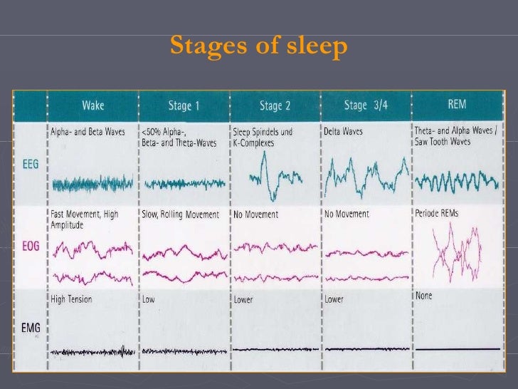
Here is a summary of how the "big three" measurements define each stage:
-
Stage W (Wakefulness):
- EEG: Shows beta waves when alert with eyes open, and alpha waves when relaxed with eyes closed.
- EOG: Shows blinking and normal, rapid eye movements.
- EMG: High muscle tone.
-
Stage N1 (NREM 1 - Light Sleep): This is the brief transition into sleep.
- EEG: Alpha waves disappear and are replaced by low-amplitude theta waves.
- EOG: Slow, rolling eye movements begin.
- EMG: Muscle tone is slightly reduced compared to wake.
- You might experience hypnic jerks (sudden muscle twitches) in this stage.
-
Stage N2 (NREM 2 - True Sleep): You spend the most time here (~50% of the night).
- EEG: Continues to show theta waves but is defined by two unique features: sleep spindles (brief bursts of fast activity) and K-complexes (large, slow waves).
- EOG: Eye movements stop.
- EMG: Muscle tone continues to decrease.
-
Stage N3 (NREM 3 - Deep Sleep / Slow-Wave Sleep): The most restorative stage.
- EEG: Dominated by high-amplitude, slow delta waves. An epoch is scored as N3 when more than 20% of it consists of these waves.
- EOG: No eye movements.
- EMG: Low muscle tone. It's very difficult to be awakened from this stage.
-
Stage R (REM Sleep - Paradoxical Sleep): The stage most associated with vivid dreaming.
- EEG: Brain activity becomes very active again, looking similar to wakefulness with low-amplitude, mixed-frequency waves.
- EOG: Shows bursts of rapid eye movements.
- EMG: This is the key differentiator. The body's voluntary muscles are essentially paralyzed, a state called atonia. This prevents you from acting out your dreams.
- This stage is called "paradoxical" because the brain is highly active, while the body is immobile.
Test your understanding!
A sleep scientist is looking at a 30-second epoch of a polysomnogram. They observe the following:
- EEG: Low-amplitude, mixed-frequency brain waves, looking very similar to the awake state.
- EOG: Bursts of horizontal, jerky movements.
- EMG: The signal is almost completely flat.
Which stage of sleep is this person in?
Show answer
This person is in Stage R (REM sleep). The combination of an active, "awake-like" EEG, rapid eye movements (EOG), and muscle paralysis (EMG atonia) are the defining features of this stage.
Conclusion
In this lesson, we have laid the crucial scientific groundwork for our exploration of sleep's architecture. We moved from the abstract concept of sleep to the concrete, measurable reality captured by technology.
Key Takeaways:
- Polysomnography (PSG) is the scientific method used to record multiple physiological signals during sleep.
- Sleep staging primarily relies on analyzing the EEG (brain waves), EOG (eye movements), and EMG (muscle tension).
- Sleep scientists score the night in 30-second epochs, assigning each a stage based on the AASM scoring manual.
- The stages are Wake (W), three non-rapid eye movement stages (N1, N2, N3), and one rapid eye movement stage (R).
- Each stage is defined by a unique signature, such as the delta waves of N3 deep sleep or the combination of a fast EEG and muscle atonia in REM sleep.
Preview of the Next Lesson:
Now that we have learned how to identify the different stages of sleep, we can begin to explore their individual characteristics and functions. In the next lesson, we will take a closer look at the non-rapid eye movement (NREM) stages: N1, N2, and the deeply restorative N3, or slow-wave sleep. We will examine what the brain and body are doing during these crucial periods of rest.